 뒤로
뒤로Blood: Structure, Function, and Clinical Significance
스터디 가이드 - 스마트 노트
 자료에 맞춘 맞춤형 노트, 핵심 정의, 예시, 맥락을 확장해 제공합니다.
Blood: Structure, Function, and Clinical Significance
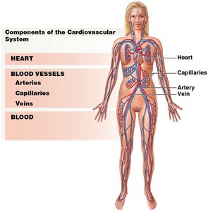
Overview of the Cardiovascular System
The cardiovascular system is essential for transporting substances throughout the body and maintaining homeostasis. It consists of the heart (a muscular pump), blood vessels (arteries, veins, and capillaries), and blood (a fluid connective tissue).
Heart: Pumps blood throughout the body.
Blood Vessels: Arteries carry blood away from the heart, veins return blood to the heart, and capillaries allow exchange with tissues.
Blood: Transports gases, nutrients, wastes, and hormones; regulates pH and temperature; defends against pathogens; and restricts fluid loss.
Physical Characteristics and Composition of Blood
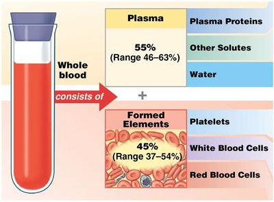
Blood is a specialized connective tissue composed of plasma and formed elements. It is slightly alkaline (pH 7.35–7.45), has high viscosity, and constitutes about 7% of body weight.
Volume: 5–6 L in adult males, 4–5 L in adult females.
Temperature: 38°C (100.4°F).
Plasma
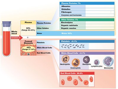
Plasma makes up about 55% of blood volume and is primarily water (over 90%), with dissolved proteins, electrolytes, nutrients, and wastes.
Plasma Proteins: Albumins (osmotic pressure, transport), globulins (antibodies, transport), fibrinogen (clotting), and others.
Origins: Most plasma proteins are synthesized in the liver; antibodies are produced by plasma cells.
Formed Elements
Formed elements constitute about 45% of blood and include red blood cells (erythrocytes), white blood cells (leukocytes), and platelets (thrombocytes).
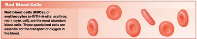
Red Blood Cells (RBCs): 99% of formed elements; transport oxygen and carbon dioxide.
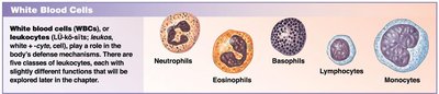
White Blood Cells (WBCs): Immune defense; several types with specialized functions.
Platelets: Cell fragments involved in blood clotting.

Red Blood Cells (Erythrocytes)
Structure and Function
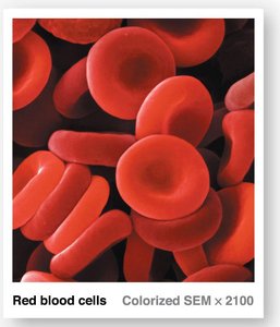
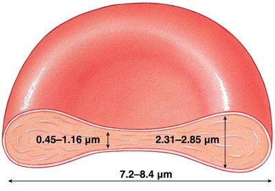
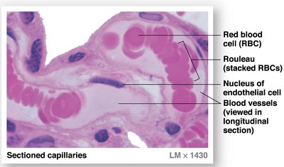
Red blood cells are small, biconcave discs lacking nuclei and most organelles, optimized for gas transport. Their shape increases surface area for gas exchange and allows flexibility in capillaries.
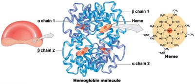
Hemoglobin: The protein responsible for oxygen and carbon dioxide transport. Each molecule has four subunits, each with a heme group containing iron.
Normal Counts: Males: 4.5–6.3 million/μL; Females: 4.2–5.5 million/μL.
Hematocrit: Percentage of blood volume occupied by RBCs (Males: 46%, Females: 42%).
RBC Life Cycle and Erythropoiesis
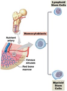
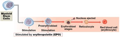
RBCs live about 120 days. Erythropoiesis (RBC production) occurs in red bone marrow and is stimulated by erythropoietin (EPO), mainly from the kidneys in response to hypoxia.
Hemopoiesis: Formation of all blood cells from hematopoietic stem cells.
Recycling: Macrophages in the spleen, liver, and bone marrow break down old RBCs; iron is recycled, and heme is converted to bilirubin.
Blood Types and Transfusion Compatibility
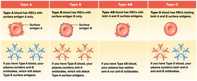
Blood types are determined by surface antigens (A, B, Rh) on RBCs. The presence or absence of these antigens defines the ABO and Rh blood groups, which are critical for safe transfusions.
Agglutinogens: Surface antigens on RBCs.
Agglutinins: Plasma antibodies that react with foreign antigens, causing agglutination (clumping).
Universal Donor: Type O−; Universal Recipient: Type AB+.
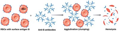
Hemolytic Disease of the Newborn (HDN)
HDN occurs when an Rh− mother carries an Rh+ fetus, leading to maternal antibody production that can attack fetal RBCs in subsequent pregnancies. Prevention involves administering RhoGAM to the mother.
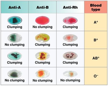
White Blood Cells (Leukocytes)
Types and Functions
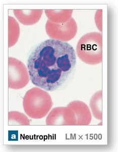
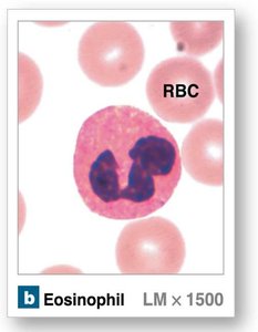
WBCs are crucial for immune defense and are classified as granulocytes or agranulocytes. They can migrate into tissues and respond to infection, inflammation, and injury.
Neutrophils: Phagocytize bacteria; first responders to infection.
Eosinophils: Attack parasites, modulate allergic responses.
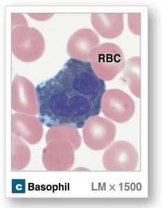
Basophils: Release histamine and heparin; involved in inflammation and allergy.
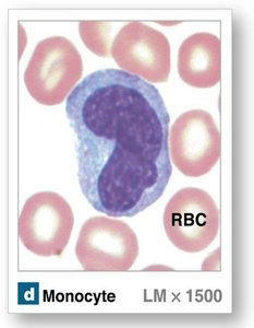
Monocytes: Become macrophages in tissues; phagocytize pathogens and debris.
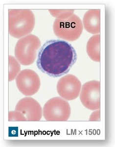
Lymphocytes: T cells, B cells, and NK cells; adaptive immunity.
WBC Disorders
Leukopenia: Low WBC count.
Leukocytosis: High WBC count.
Leukemia: Cancer of blood-forming tissues, characterized by abnormal WBCs.
Platelets (Thrombocytes)
Structure and Function
Platelets are small cell fragments essential for hemostasis (stopping blood loss). They circulate for 9–12 days and are stored in the spleen and other organs.
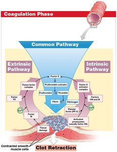
Hemostasis: Involves vascular, platelet, and coagulation phases to form a stable blood clot.
Clot Retraction: Platelets contract to reduce the size of the damaged area and facilitate repair.
Anticoagulants: Substances like antithrombin-III and heparin prevent excessive clotting.

Blood Disorders and Clinical Considerations
Nutritional and Congenital Blood Disorders
Iron Deficiency Anemia: Low iron impairs hemoglobin synthesis.
Pernicious Anemia: Vitamin B12 deficiency affects RBC production.
Sickle Cell Anemia: Genetic disorder causing abnormal hemoglobin and RBC shape.
Hemophilia: Inherited deficiency of clotting factors.
Thalassemias: Inability to produce functional hemoglobin subunits.
Blood Infections and Cancers
Bacteremia: Bacteria in the blood.
Sepsis: Systemic infection with multiplying bacteria and toxins.
Viremia: Viruses in the blood.
Malaria: Parasitic infection of RBCs by Plasmodium.
Leukemias: Cancers of blood-forming tissues, classified as myeloid or lymphoid.
Key Equations and Tables
Blood Volume Calculation:
Summary Table: Major Blood Cell Types
Cell Type | Main Function | Relative Abundance |
|---|---|---|
Red Blood Cell (Erythrocyte) | Oxygen and CO2 transport | ~99% of formed elements |
White Blood Cell (Leukocyte) | Immune defense | <1% of formed elements |
Platelet (Thrombocyte) | Clotting | <1% of formed elements |
Summary Table: ABO Blood Types and Antibodies
Blood Type | Surface Antigen(s) | Plasma Antibody(ies) |
|---|---|---|
A | A | Anti-B |
B | B | Anti-A |
AB | A and B | None |
O | None | Anti-A, Anti-B |
Additional info: This guide integrates foundational concepts, clinical relevance, and visual aids to support mastery of blood structure and function for Anatomy & Physiology students.
