 뒤로
뒤로Blood: Structure, Function, and Disorders – Anatomy & Physiology Study Notes
스터디 가이드 - 스마트 노트
 자료에 맞춘 맞춤형 노트, 핵심 정의, 예시, 맥락을 확장해 제공합니다.
Blood: Structure, Function, and Disorders
Introduction to Blood
Blood is a specialized connective tissue that plays a vital role in the maintenance of homeostasis within the human body. It is the only fluid tissue in the body and consists of a liquid matrix called plasma in which various cells and cell fragments (formed elements) are suspended.
Functions of Blood
Transportation
Oxygen and Nutrients: Blood delivers oxygen from the lungs and nutrients from the digestive tract to all body cells.
Waste Removal: Blood transports metabolic waste products from cells to elimination sites (lungs for CO2, kidneys for nitrogenous wastes).
Hormone Transport: Blood carries hormones from endocrine organs to target tissues.
Regulation
Body Temperature: Blood absorbs and distributes heat throughout the body.
pH Balance: Blood maintains normal pH using buffers and by transporting acids/bases.
Fluid Volume: Blood proteins prevent excessive fluid loss from the bloodstream.
Protection
Clot Formation: Platelets and plasma proteins initiate clotting to prevent blood loss.
Immunity: White blood cells (leukocytes) and antibodies defend against pathogens.
Composition of Blood
Blood is composed of plasma (the liquid matrix) and formed elements (cells and cell fragments).
Plasma: The non-living fluid matrix, making up about 55% of whole blood.
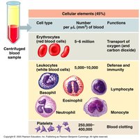
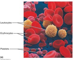
Formed Elements: Includes erythrocytes (red blood cells), leukocytes (white blood cells), and platelets (cell fragments).
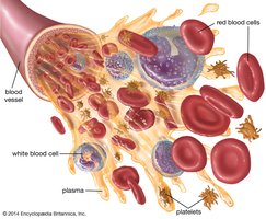
Major Components of Whole Blood
Hematocrit: The percentage of blood volume occupied by red blood cells. Normal values: Males 47% ± 5%, Females 42% ± 5%.
Physical Characteristics: Color varies with oxygen content (scarlet red when oxygen-rich, dark red when oxygen-poor). Blood pH is 7.35–7.45. Average adult blood volume is ~5 liters, about 8% of body weight.
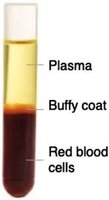
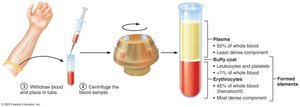
Blood Plasma
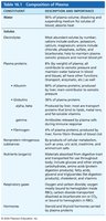
Plasma is a straw-colored, sticky fluid composed mostly of water (about 90%) and contains dissolved solutes such as nutrients, gases, hormones, wastes, proteins, and electrolytes.
Plasma Proteins: Albumin (maintains osmotic pressure), globulins (immune function), fibrinogen (clotting).
Other Solutes: Electrolytes, nutrients, respiratory gases, hormones, and waste products.
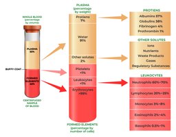
Formed Elements
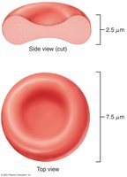
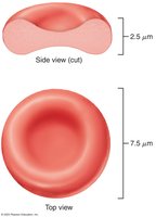
Erythrocytes (RBCs): Anucleate, biconcave discs specialized for oxygen transport.
Leukocytes (WBCs): True cells involved in immune defense.
Platelets: Cell fragments essential for blood clotting.
Erythrocytes: Structure and Function
Structural Characteristics
Biconcave Disc Shape: Maximizes surface area for gas exchange and allows flexibility to pass through capillaries.
Anucleate and Lacking Organelles: Increases space for hemoglobin; rely on anaerobic metabolism.
Life Span: 100–120 days.
Function: Gas Transport
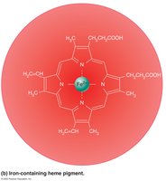
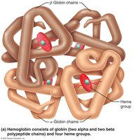
Hemoglobin (Hb): The protein responsible for oxygen and carbon dioxide transport. Each Hb molecule consists of four polypeptide chains (two alpha, two beta) and four heme groups, each containing an iron atom that binds O2.
Oxygen Binding: Each Hb can bind up to four O2 molecules.
CO2 Transport: About 20% of CO2 binds to Hb (carbaminohemoglobin).
Oxygen Loading and Unloading
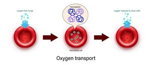
In Lungs: O2 binds to Hb, forming oxyhemoglobin (bright red).
In Tissues: O2 is released, forming deoxyhemoglobin (dark red).
CO2 Loading: CO2 binds to Hb in tissues, forming carbaminohemoglobin.
Hematopoiesis: Formation of Blood Cells
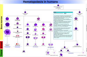
Hematopoiesis is the process of blood cell formation, occurring primarily in red bone marrow. All formed elements arise from hematopoietic stem cells (hemocytoblasts).
Stem Cells: Hemocytoblasts differentiate into various blood cell types.
Location: Red bone marrow of axial skeleton, girdles, and proximal epiphyses of humerus and femur.
Erythropoiesis: Formation of Erythrocytes
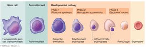
Stages: Hematopoietic stem cell → myeloid stem cell → proerythroblast → basophilic erythroblast → polychromatic erythroblast → orthochromatic erythroblast → reticulocyte → erythrocyte.
Time Frame: Approximately 15 days from stem cell to mature RBC.
Regulation of Erythropoiesis
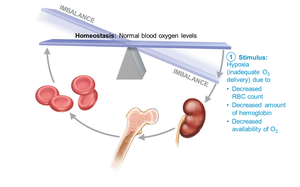
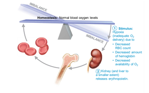
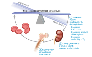
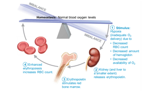
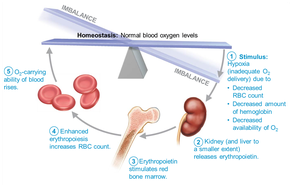
Homeostasis: Balance between RBC production and destruction is crucial. Too few RBCs cause hypoxia; too many increase blood viscosity.
Hormonal Control: Erythropoietin (EPO), produced by the kidneys in response to hypoxia, stimulates RBC production.
Dietary Requirements: Iron, amino acids, vitamin B12, and folic acid are essential for erythropoiesis.
Life Cycle and Fate of Erythrocytes
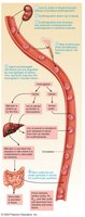
Destruction: Old RBCs are engulfed by macrophages in the spleen, liver, and bone marrow.
Iron Recycling: Iron is salvaged, stored as ferritin or hemosiderin, and transported by transferrin.
Heme Degradation: Heme is degraded to bilirubin, which is excreted in bile.
Erythrocyte Disorders
Anemia
Anemia is a condition in which the blood has an abnormally low oxygen-carrying capacity, insufficient to support normal metabolism. Causes include blood loss, decreased RBC production, or increased RBC destruction.
Sickle-Cell Anemia
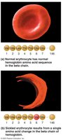
Sickle-cell anemia is a genetic disorder resulting from a single amino acid change in the beta chain of hemoglobin, causing RBCs to become sickle-shaped under low oxygen conditions. This leads to blockages in small blood vessels and reduced oxygen delivery.
Polycythemia
Polycythemia is an abnormal excess of RBCs, increasing blood viscosity and risk of clotting. Causes include bone marrow cancer or artificial means such as blood doping.
Summary Table: Major Components of Blood
Component | Percentage | Main Function |
|---|---|---|
Plasma | ~55% | Transport of nutrients, wastes, hormones; maintains blood volume and pH |
Erythrocytes (RBCs) | ~45% | Transport of oxygen and carbon dioxide |
Leukocytes (WBCs) | <1% | Defense and immunity |
Platelets | <1% | Blood clotting |
Additional info: These notes are based on Chapter 16 (Blood) of a standard Anatomy & Physiology textbook and are suitable for college-level study and exam preparation.
