 뒤로
뒤로Bones and Bone Tissue: Structure, Function, Growth, and Repair
스터디 가이드 - 스마트 노트
 자료에 맞춘 맞춤형 노트, 핵심 정의, 예시, 맥락을 확장해 제공합니다.
Bones and Bone Tissue
The Skeletal System: Overview
The skeletal system is a complex organ system that includes bones, joints, and supporting tissues. Bones are the main organs, with adults typically having 206 bones. Each bone contains osseous tissue, dense regular and irregular connective tissue, and bone marrow.
Bones: Provide structure and support.
Joints: Allow movement and flexibility.
Bone Marrow: Site of blood cell formation and fat storage.
Functions of the Skeletal System
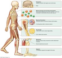
The skeletal system performs several essential functions for the human body:
Protection: Bones such as the skull, sternum, and ribs protect vital organs.
Mineral Storage and Acid-Base Homeostasis: Bones store minerals (calcium, phosphorus, magnesium) critical for electrolyte and acid-base balance.
Blood Cell Formation: Red bone marrow is the site of hematopoiesis (formation of blood cells).
Fat Storage: Yellow bone marrow stores triglycerides in adipocytes.
Movement: Bones serve as attachment sites for muscles, enabling movement.
Support: The skeleton supports body weight and provides structural framework.
Bone Structure
Classification of Bones by Shape
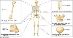
Bones are classified based on their shape, which relates to their function and location:
Long Bones: Longer than wide (e.g., humerus, femur).
Short Bones: Cube-shaped, as long as wide (e.g., wrist, ankle bones).
Flat Bones: Thin and broad (e.g., skull, pelvis).
Irregular Bones: Irregular shapes (e.g., vertebrae).
Sesamoid Bones: Small, flat, oval-shaped, within tendons (e.g., patella).
Structure of a Long Bone
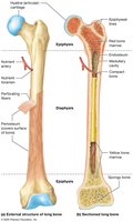
Long bones have a specialized structure to support their function:
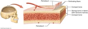
Periosteum: Outer membrane with blood vessels and nerves.
Perforating Fibers: Collagen anchors attaching periosteum to bone.
Diaphysis: Shaft containing medullary cavity lined by endosteum and filled with marrow.
Epiphyses: Ends of the bone, filled with red marrow and covered with articular cartilage.
Compact Bone: Dense outer layer resisting compression and twisting.
Spongy Bone: Inner honeycomb-like structure housing bone marrow.
Epiphyseal Lines: Remnants of growth plates.
Structure of Short, Flat, Irregular, and Sesamoid Bones
These bones share similarities with long bones but have fewer structures. In flat bones, spongy bone is called diploë, and some skull bones contain sinuses to reduce weight.
Blood and Nerve Supply to Bone
Bones are highly vascularized and innervated:
Short, flat, irregular, and sesamoid bones receive blood from periosteal vessels.
Long bones are supplied by periosteal vessels and nutrient arteries entering through the nutrient foramen.
Red and Yellow Marrow
Bone marrow exists in two forms:
Red Bone Marrow: Hematopoietic tissue, abundant in children, limited to certain bones in adults.
Yellow Bone Marrow: Contains adipocytes and blood vessels, predominant in adults.
Bone Marrow Transplantation
Bone Marrow Transplantation
Used to treat diseases like leukemia and sickle-cell anemia. Donor marrow is harvested and transplanted after recipient’s marrow is destroyed. Peripheral Blood Stem Cell (PBSC) donation is an alternative, where stem cells are collected from blood after stimulation.
The Extracellular Matrix of Bone
Inorganic Matrix
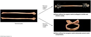
Comprises about 65% of bone weight, mainly hydroxyapatite crystals (calcium and phosphorus), providing strength and resistance to compression. Other ions include bicarbonate, potassium, magnesium, and sodium.
Organic Matrix (Osteoid)
About 35% of bone weight, consists of collagen fibers, proteoglycans, glycosaminoglycans, glycoproteins, and osteocalcin. Collagen resists torsion and tensile forces, while osteocalcin organizes the inorganic matrix.
Bone Cells
Types of Bone Cells
Bone is dynamic, constantly remodeled by three main cell types:
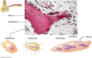
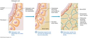
Osteoblasts: Build bone, derived from osteogenic cells, perform bone deposition.
Osteocytes: Mature osteoblasts trapped in lacunae, maintain ECM.
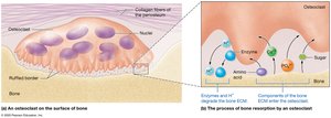
Osteoclasts: Large, multinucleated cells that break down bone (bone resorption).
Osteopetrosis
"Marble Bone Disease" caused by defective osteoclasts, leading to increased bone mass but weak, brittle bones. Infantile form is severe and can be fatal; adult form causes pain and fractures.
Histology of Bone
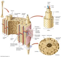
Compact Bone
Hard, dense outer shell composed of osteons (Haversian systems):
Lamellae: Concentric rings of bone.
Central Canal: Contains blood vessels and nerves.
Lacunae: Small cavities housing osteocytes.
Canaliculi: Tiny canals connecting lacunae.
Interstitial and Circumferential Lamellae: Strengthen bone.
Perforating Canals: Connect central canals and carry blood vessels.
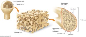
Spongy Bone
Consists of trabeculae, covered with endosteum, containing lamellae, lacunae, and canaliculi but lacking central canals. Blood supply comes from bone marrow vessels.
Bone Formation: Ossification
Ossification (Osteogenesis)
Process of bone formation, continuing through childhood. Two types:
Primary (Woven) Bone: Immature, irregular collagen, abundant osteocytes, little inorganic matrix.
Secondary (Lamellar) Bone: Mature, organized lamellae, parallel collagen, more inorganic matrix.
Types of Ossification
Intramembranous Ossification: Forms flat bones (skull, clavicles) from mesenchymal membrane.
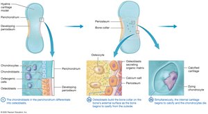
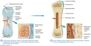
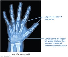
Endochondral Ossification: Forms long and short bones from hyaline cartilage model.
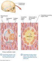
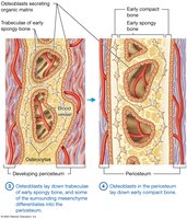
Steps of Intramembranous Ossification
Osteoblasts develop in primary ossification center from mesenchymal cells.
Osteoblasts secrete organic matrix, which calcifies; trapped osteoblasts become osteocytes.
Osteoblasts lay down trabeculae of early spongy bone; some mesenchyme becomes periosteum.
Osteoblasts in periosteum lay down early compact bone; matrix is remodeled.
Steps of Endochondral Ossification
Chondroblasts in perichondrium differentiate into osteoblasts.
Osteoblasts build bone collar on external surface; internal cartilage calcifies and chondrocytes die.
Osteoblasts replace calcified cartilage with early spongy bone; secondary ossification centers and medullary cavity develop.
Medullary cavity enlarges; remaining cartilage replaced by bone; epiphyses finish ossifying.
Comparison: Intramembranous vs. Endochondral Ossification
Flat bones form by intramembranous ossification; long and short bones by endochondral ossification.
Order of bone formation differs: compact bone forms first in endochondral, spongy bone first in intramembranous.
Bone Growth
Longitudinal Growth
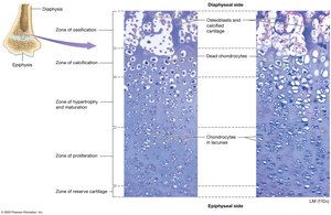
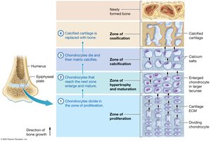
Occurs at the epiphyseal plate, which has five zones:
Zone of Reserve Cartilage: Cells not directly involved in growth.
Zone of Proliferation: Actively dividing chondrocytes.
Zone of Hypertrophy and Maturation: Mature chondrocytes.
Zone of Calcification: Dead, calcified chondrocytes.
Zone of Ossification: Calcified chondrocytes and osteoblasts build bone.
Appositional Growth
Growth in bone width occurs as osteoblasts lay down new circumferential lamellae, thickening the compact bone and enlarging the medullary cavity.
The Role of Hormones in Bone Growth
Growth Hormone: Increases mitosis of chondrocytes, activity of osteogenic cells, and stimulates osteoblasts.
Testosterone: Promotes appositional growth and mitosis, accelerates epiphyseal plate closure.
Estrogen: Similar effects, but less pronounced; epiphyseal plates close earlier in females.
Gigantism and Acromegaly
Excess growth hormone causes gigantism (before plate closure) or acromegaly (after closure), leading to abnormal bone and tissue growth.

Bone Remodeling
Bone Remodeling Process
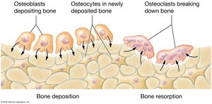
Bone remodeling is a continuous process involving bone deposition (by osteoblasts) and bone resorption (by osteoclasts). It maintains calcium homeostasis, repairs bone, replaces old bone, and adapts to stress.
Bone Deposition
Osteoblasts secrete organic matrix and facilitate inorganic matrix formation.
Calcification occurs as calcium ions crystallize and vesicles rupture.
Bone Resorption
Osteoclasts secrete hydrogen ions and enzymes to break down bone matrix.
Minerals and organic components are released for use elsewhere in the body.
Bone Remodeling in Response to Tension and Stress
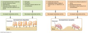
Compression and tension stimulate bone deposition.
Continuous pressure stimulates bone resorption.
Other Factors Influencing Bone Remodeling
Hormones: Testosterone promotes deposition; estrogen inhibits osteoclasts.
Age: Hormone levels decline, reducing bone remodeling.
Nutrient Intake: Calcium, vitamin D, K, C, and protein are essential for bone health.
Calcium Ion Intake and Fracture Risk
Recommended intake is 700–1,200 mg/day, but excessive calcium does not necessarily reduce fracture risk and may have adverse effects.
Bone Remodeling and Calcium Ion Homeostasis
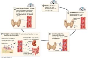
Calcium ions are vital for muscle contraction, nerve transmission, and blood clotting. Blood calcium is regulated by:
Parathyroid Hormone (PTH): Increases blood calcium.
Calcitonin: Decreases blood calcium (less potent in adults).
Bone Repair
Steps of Fracture Healing
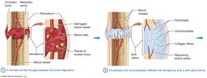
Hematoma fills the gap between bone fragments.
Fibroblasts and chondroblasts infiltrate hematoma, forming a soft callus.
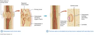
Osteoblasts build a bone callus.
Bone callus is remodeled; primary bone replaced with secondary bone.
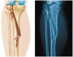
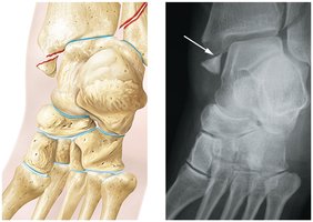
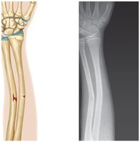
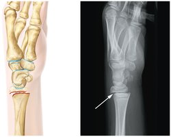
Classes and Types of Fractures
Simple (Closed) Fractures: Skin and tissue remain intact.
Compound (Open) Fractures: Damage to surrounding tissue.
Treatment: Stabilization and immobilization; closed or open reduction.
Table: Types of Fractures
Type | Description |
|---|---|
Transverse | Fracture perpendicular to bone's axis |
Spiral | Fracture spirals around bone |
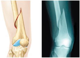
Comminuted | Bone shattered into multiple fragments |
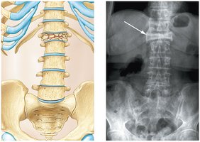
Compression | Bone crushed under weight |
Greenstick | Bone bends and partially breaks (common in children) |
Epiphyseal | Fracture at epiphyseal plate |
Additional info: Other types may include oblique, impacted, and pathologic fractures. |
Summary
The skeletal system is essential for protection, movement, support, mineral storage, and blood cell formation. Bone structure, growth, remodeling, and repair are regulated by cellular activity, hormones, and nutrients. Understanding these processes is fundamental for Anatomy & Physiology students.
