 뒤로
뒤로Cell Cycle, DNA Replication, and Protein Synthesis: Study Notes for Anatomy & Physiology
스터디 가이드 - 스마트 노트
 자료에 맞춘 맞춤형 노트, 핵심 정의, 예시, 맥락을 확장해 제공합니다.
Cells: The Living Units
Cell Cycle
The cell cycle describes the series of events that cells undergo from their formation to their division. It is essential for growth, maintenance, and repair in multicellular organisms.
Interphase: The period between cell formation and cell division, where the cell grows and performs its functions. Nuclear material is called chromatin.
Mitotic Phase: The phase where the cell divides into two daughter cells.
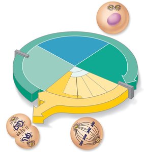
Interphase Subphases
G1 (Gap 1): Vigorous growth and metabolism. Cells that permanently cease dividing are said to be in the G0 phase.
S (Synthetic): DNA replication occurs.
G2 (Gap 2): Preparation for cell division.
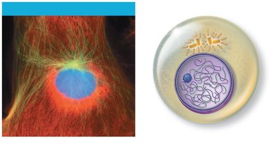
DNA Replication
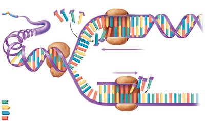
DNA replication is the process by which a cell copies its DNA before division, ensuring each daughter cell receives a complete set of genetic instructions.
Replication Bubbles: DNA helices are separated, forming bubbles with replication forks at each end.
DNA Polymerase: Adds nucleotides at the RNA primer, synthesizing one leading strand continuously and one lagging strand discontinuously.
DNA Ligase: Splices short segments of the lagging strand together.
Semiconservative Replication: Each new DNA molecule consists of one old and one new strand.
Cell Division
Cell division is crucial for growth, tissue repair, and reproduction. There are two main types:
Meiosis: Produces gametes (sperm and egg cells).
Mitotic Cell Division: Produces clones for body growth and tissue repair. Occurs continuously in some cells (e.g., skin, intestinal lining), but not in most mature nervous, skeletal, or cardiac muscle cells.
Events of Cell Division
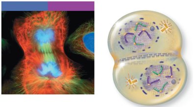
Mitosis: Division of the nucleus, ensuring each cell receives a copy of replicated DNA. Four stages: Prophase, Metaphase, Anaphase, Telophase.
Cytokinesis: Division of cytoplasm by cleavage furrow.
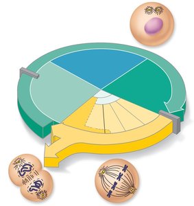
Mitosis Stages
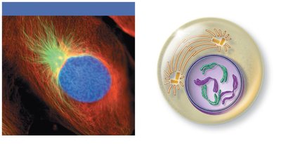
Prophase: Chromosomes become visible, centrosomes migrate, mitotic spindles form, nuclear envelope fragments.
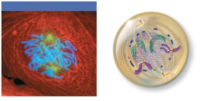
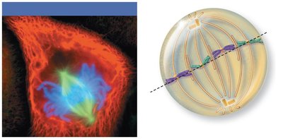
Metaphase: Chromosomes align at the cell equator (metaphase plate).
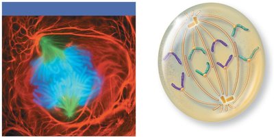
Anaphase: Centromeres split, chromatids become chromosomes, pulled toward poles.
Telophase: Chromosomes uncoil to form chromatin, new nuclear membranes form, nucleoli reappear, spindle disappears.
Control of Cell Division
Cell division is tightly regulated by internal and external signals.
"Go" Signals: Critical cell volume, growth factors, hormones, and contact inhibition.
Cyclins and Cdks: Regulatory proteins that accumulate during interphase and activate enzymes for division.
Checkpoints: G1 checkpoint (restriction point) is most important; G2 checkpoint requires MPF (M-phase promoting factor).
Repressor Genes: Inhibit cell division (e.g., P53 gene).
Protein Synthesis
DNA and Genes
DNA is the master blueprint for protein synthesis. A gene is a segment of DNA coding for one polypeptide. DNA bases are A (adenine), G (guanine), T (thymine), and C (cytosine).
Triplets: Three sequential DNA bases specify the coding for amino acids.
Exons: Coding segments of genes.
Introns: Noncoding segments.
Role of RNA
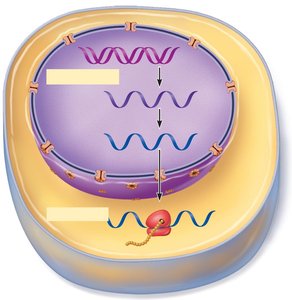
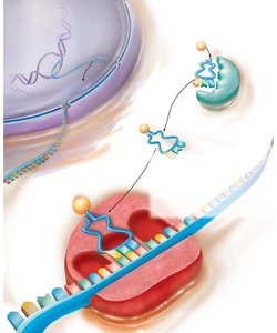
Messenger RNA (mRNA): Carries instructions from DNA to ribosomes.
Ribosomal RNA (rRNA): Structural component of ribosomes, helps translate mRNA.
Transfer RNA (tRNA): Binds amino acids and pairs with mRNA codons at ribosome.
Steps of Protein Synthesis
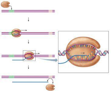
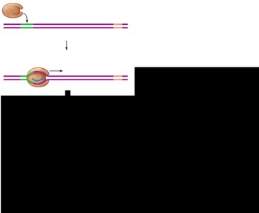
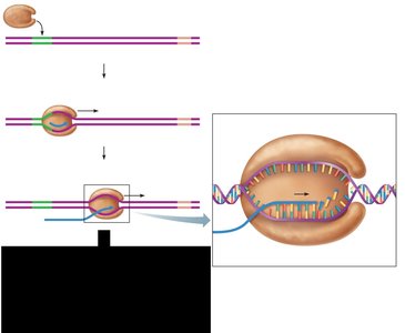
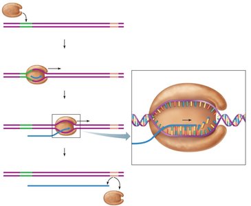
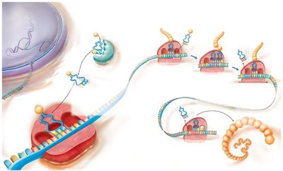
Transcription: DNA information is coded in mRNA. Occurs in three phases:
Initiation: RNA polymerase binds to promoter, separates DNA strands.
Elongation: RNA polymerase adds complementary nucleotides.
Termination: RNA polymerase releases completed mRNA transcript.
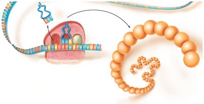
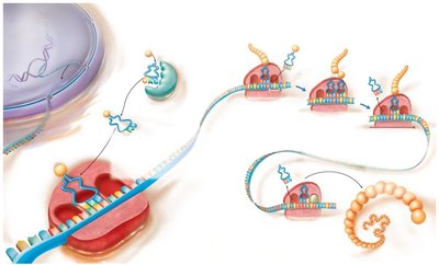
Translation: mRNA is decoded to assemble polypeptides. Involves mRNAs, tRNAs, and rRNAs.
Genetic Code
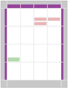
The genetic code is based on three-base sequences (codons) on mRNA, each specifying an amino acid. Some amino acids are represented by more than one codon.
Translation: Sequence of Events
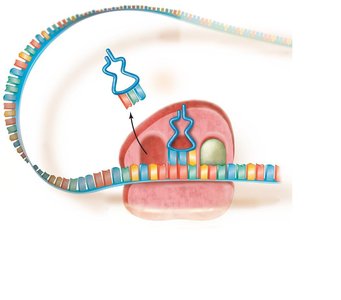
Initiation: Small ribosomal subunit binds to initiator tRNA and mRNA, scans for start codon. Large subunit attaches, forming functional ribosome.
Elongation:
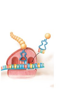
Codon Recognition: tRNA binds complementary codon in A site.
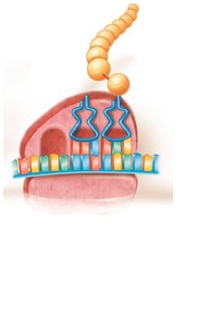
Peptide Bond Formation: Amino acid in P site is bonded to amino acid in A site.
Translocation: tRNAs move one position: A → P; P → E.
Termination: Stop codon enters A site, release factor binds, polypeptide released, ribosome subunits separate.
Polyribosomes
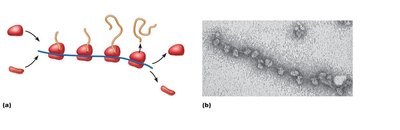
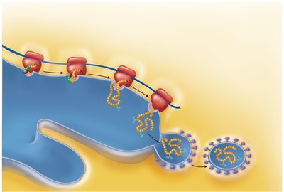
A polyribosome is a complex of multiple ribosomes reading a single mRNA strand, producing multiple copies of the same protein.
Role of Rough ER in Protein Synthesis
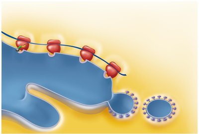
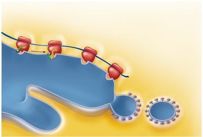
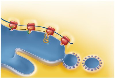
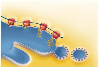
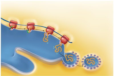
The rough endoplasmic reticulum (ER) is involved in processing proteins synthesized by ribosomes. The mRNA–ribosome complex is directed to the rough ER by a signal-recognition particle (SRP), where the protein enters the ER, may be modified, and is enclosed in a vesicle for transport to the Golgi apparatus.
Summary: From DNA to Proteins
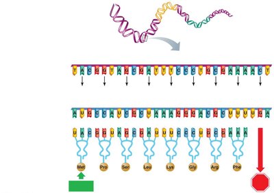
Complementary base pairing directs transfer of genetic information from DNA to protein.
DNA triplets → mRNA codons → tRNA anticodons → amino acid sequence.
Anticodon sequence is identical to DNA sequence except uracil replaces thymine.
Other Roles of DNA and Cell Regulation
Noncoding DNA Functions
Antisense RNA: Prevents translation of protein-coding RNA.
MicroRNA: Silences mRNAs from certain exons.
Riboswitches: Folded RNAs acting as switches to regulate protein synthesis.
Cytosolic Protein Degradation
Autophagy: Degradation of cytoplasmic bits and organelles by lysosomes.
Ubiquitins: Tag damaged or unneeded proteins for digestion by proteasomes.
Extracellular Materials
Body Fluids: Interstitial fluid, blood plasma, cerebrospinal fluid.
Cellular Secretions: Intestinal and gastric fluids, saliva, mucus, serous fluids.
Extracellular Matrix: Jellylike mesh of proteins and polysaccharides acting as "glue" to hold cells together.
Developmental Aspects of Cells
All cells contain the same DNA, but chemical signals channel cells into specific developmental pathways (cell differentiation).
Apoptosis and Cell Division Rates
Apoptosis: Programmed cell death, important for eliminating excess cells during development.
Hyperplasia: Increase in cell numbers when needed.
Atrophy: Decrease in cell size due to loss of stimulation or use.
Theories of Cell Aging
Wear and Tear Theory: Chemical insults and free radicals accumulate over time.
Mitochondrial Theory: Free radicals in mitochondria reduce energy production.
Immune System Disorders: Autoimmune responses and weakened immunity contribute to aging.
Genetic Theory: Cessation of mitosis and cell aging programmed into genes. Telomeres may determine cell division limits; telomerase lengthens telomeres in germ cells.
