 뒤로
뒤로Cryder Chapter 3 practice
스터디 가이드 - 스마트 노트
 자료에 맞춘 맞춤형 노트, 핵심 정의, 예시, 맥락을 확장해 제공합니다.
Plasma Membrane Structure
Fluid Mosaic Model
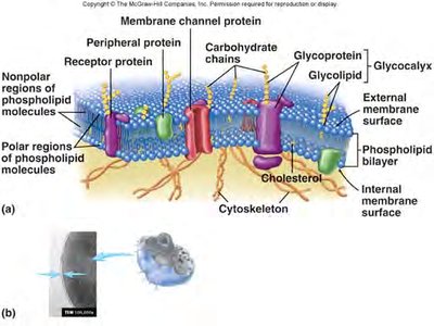
The plasma membrane is a dynamic structure that encloses the cell, separating the internal cytoplasm and nucleus from the external environment. It is described by the fluid mosaic model, which highlights its flexible, mosaic-like arrangement of molecules.
Phospholipid Bilayer: Composed of two layers of phospholipids with hydrophilic (water-attracting) heads facing outward and hydrophobic (water-repelling) tails facing inward, creating a semi-permeable barrier.
Proteins:
Peripheral Proteins: Attached to the inner or outer surface of the membrane, involved in signaling and maintaining cell shape.
Integral Proteins: Span the membrane, functioning as channels, carriers, or receptors.
Channel Proteins: Form passageways for specific molecules or ions.
Cholesterol: Interspersed within the bilayer, stabilizes membrane fluidity and structure.
Carbohydrates: Attached to proteins (glycoproteins) or lipids (glycolipids), forming the glycocalyx for cell recognition and adhesion.
Plasma Membrane Specializations
Microvilli: Finger-like projections that increase surface area for absorption, especially in the kidney and intestine.
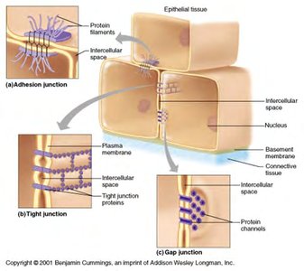
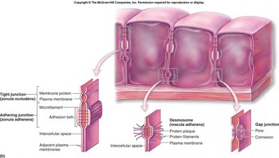
Membrane Junctions: Specialized connections between adjacent cells:
Tight Junctions: Create impermeable barriers to prevent leakage between cells.
Desmosomes: Provide strong adhesion between cells, maintaining tissue integrity.
Gap Junctions: Allow direct passage of ions and small molecules between cells for communication.
Plasma Membrane Function
Passive Membrane Transport
Passive transport does not require cellular energy and relies on concentration gradients.
Simple Diffusion: Movement of molecules from high to low concentration until equilibrium is reached. Small, nonpolar molecules (e.g., O2, CO2) diffuse easily.
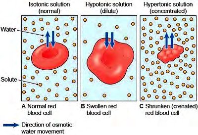
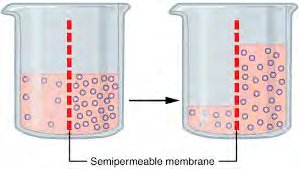
Osmosis: Diffusion of water across a selectively permeable membrane.
Hypertonic Solution: Cells lose water and shrink (crenation).
Hypotonic Solution: Cells gain water and swell or lyse.
Isotonic Solution: No net water movement; cell size remains constant.
Facilitated Diffusion: Transport of molecules via carrier proteins; specific and saturable.
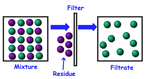
Filtration: Movement of water and solutes through a membrane due to hydrostatic pressure (e.g., formation of tissue fluid from blood capillaries).
Active Membrane Transport
Active transport requires energy (usually ATP) to move substances against their concentration gradients.
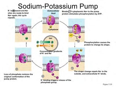
Active Transport: Uses carrier proteins (pumps) to move molecules from low to high concentration.
Na+-K+ Pump: Moves 3 Na+ ions out and 2 K+ ions into the cell, maintaining membrane potential.
Cotransport: Passive movement of one ion (e.g., Na+) powers the active transport of another molecule (e.g., glucose).
Bulk Transport:
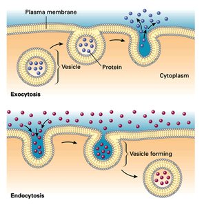
Endocytosis: Engulfment of substances into the cell via vesicles.
Pinocytosis: "Cell drinking"—uptake of liquids.
Phagocytosis: "Cell eating"—uptake of solids (e.g., debris, bacteria).
Exocytosis: Secretion of substances out of the cell via vesicles (e.g., neurotransmitter release).
Membrane Potential
Resting Membrane Potential: The inside of the cell is negatively charged relative to the outside, typically -20 to -200 mV.
Polarization: Maintained by active transport (Na+-K+ pump) and selective ion diffusion.
Cell Interactions
Glycocalyx: Carbohydrate-rich area on the cell surface, important for cell recognition and interaction (e.g., immune response, blood group determination).
Cell Cytoplasm and Organelles
Major Organelles and Their Functions
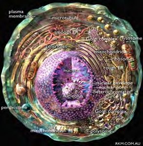
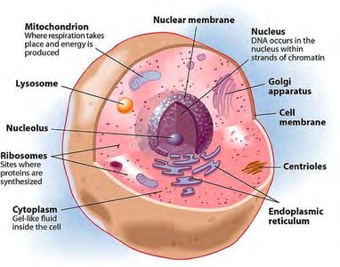
The cytoplasm contains various organelles, each with specialized functions essential for cell survival and activity.
Mitochondria: Double-membraned organelles responsible for ATP production via cellular respiration. Contain their own DNA and replicate independently.
Ribosomes: Sites of protein synthesis; can be free (cytoplasmic proteins) or membrane-bound (secretory/membrane proteins).
Endoplasmic Reticulum (ER):
Rough ER: Studded with ribosomes; synthesizes proteins.
Smooth ER: Lacks ribosomes; synthesizes lipids and detoxifies chemicals.
Golgi Apparatus: Modifies, sorts, and packages proteins and lipids for secretion or delivery to other organelles.
Lysosomes: Contain digestive enzymes for breaking down waste and cellular debris.
Vacuoles: Storage vesicles for nutrients, waste, or other substances.
Peroxisomes: Contain oxidase enzymes to detoxify harmful substances and neutralize free radicals.
Cytoskeleton: Network of protein filaments (microfilaments, intermediate filaments, microtubules) providing structural support, intracellular transport, and cell movement.
Centrioles: Organize microtubules during cell division (spindle apparatus formation).
Summary Table: Eukaryotic Cell Components
Organelle | Structure | Function |
|---|---|---|
Nucleus | Double membrane, nuclear pores | Genetic control center, stores DNA |
Ribosome | rRNA and protein, two subunits | Protein synthesis |
Rough ER | Membranous sacs with ribosomes | Protein synthesis and transport |
Smooth ER | Membranous sacs without ribosomes | Lipid synthesis, detoxification |
Golgi Apparatus | Stack of flattened sacs | Protein/lipid modification and sorting |
Lysosome | Membranous vesicle with enzymes | Digestion of macromolecules |
Peroxisome | Membranous vesicle with oxidases | Detoxification, breakdown of fatty acids |
Mitochondrion | Double membrane, cristae | ATP production |
Cytoskeleton | Protein filaments | Support, movement, transport |
Nucleus and Cell Division
Nucleus Structure and Function
Nuclear Membrane: Double-layered, with pores for material exchange.
Nucleoplasm: Fluid matrix inside the nucleus.
Nucleolus: Site of ribosome synthesis.
Chromatin: DNA-protein complex; condenses into chromosomes during cell division.
Mitotic Cell Cycle
The cell cycle consists of interphase (growth and DNA replication) and mitosis (nuclear division), followed by cytokinesis (cytoplasmic division).
Interphase:
G1 Phase: Cell growth, centriole replication.
S Phase: DNA replication (semiconservative mechanism).
G2 Phase: Preparation for division.
Mitosis:
Prophase: Chromatin condenses, nuclear envelope dissolves, centrioles migrate.
Metaphase: Chromosomes align at the cell equator.
Anaphase: Sister chromatids separate to opposite poles.
Telophase: Nuclear envelopes reform, chromosomes decondense.
Cytokinesis: Division of cytoplasm, resulting in two daughter cells.
Cancer and Cell Cycle Regulation
Neoplasm: Abnormal mass of proliferating cells.
Benign: Localized, encapsulated growths.
Malignant: Invasive, nonencapsulated, capable of metastasis (spreading to other tissues).
Protein Synthesis
Genetic Code and Protein Formation
DNA: Contains genetic instructions for protein synthesis.
Gene: Segment of DNA coding for a specific protein or RNA.
Triplet Code: Each amino acid is specified by a sequence of three nucleotides (codon).
Transcription (Nucleus)
DNA is used as a template to synthesize messenger RNA (mRNA).
mRNA is single-stranded and exits the nucleus to enter the cytoplasm.
Translation (Cytoplasm)
mRNA binds to ribosomes, where transfer RNA (tRNA) brings specific amino acids.
tRNA anticodons pair with mRNA codons, and amino acids are joined to form a polypeptide chain.
Spliceosomes and mRNA Processing
Exons: Coding regions retained in mature mRNA.
Introns: Noncoding regions removed during mRNA processing.
Degeneracy of the Genetic Code
There are 64 possible codons but only 20 amino acids, so multiple codons can code for the same amino acid.
Mutations
Substitution: One base is replaced by another.
Frame Shift: Insertion or deletion of bases alters the reading frame.
Effects range from no change to cell death or cancer.
Extracellular Materials
Types and Functions
Body Fluids: Interstitial fluid, blood plasma, cerebrospinal fluid (CSF).
Cellular Secretions: Aid in digestion (e.g., gastric fluids) and lubrication (e.g., mucus, serous fluids).
Extracellular Matrix: Network of proteins and polysaccharides providing structural support, especially in connective tissues.
Key Terms and Concepts
Phospholipid Bilayer
Integral/Peripheral Proteins
Glycocalyx
Osmosis
Facilitated Diffusion
Active Transport
Endocytosis/Exocytosis
Mitochondria
Ribosomes
Endoplasmic Reticulum
Golgi Apparatus
Lysosomes
Peroxisomes
Cytoskeleton
Centrioles
Nucleus
Chromatin/Chromosomes
Interphase/Mitosis/Cytokinesis
Mutation
Additional info: This guide expands on the provided notes with definitions, examples, and academic context to ensure completeness and clarity for Anatomy & Physiology students.
