 뒤로
뒤로Cells: The Living Units – Structure, Function, and Processes
스터디 가이드 - 스마트 노트
 자료에 맞춘 맞춤형 노트, 핵심 정의, 예시, 맥락을 확장해 제공합니다.
Cells: The Living Units
Introduction to Cells
Cells are the fundamental structural and functional units of all living organisms. The human body is composed of approximately 50 to 100 trillion cells, each specialized for particular functions. The cell theory states that all living things are made of cells, the cell is the smallest unit of life, and all cells arise from pre-existing cells.
Cell Diversity: Over 250 types of human cells exist, differing in size, shape, and subcellular components, which determine their specialized functions.
Examples: Epithelial cells (lining and transport), muscle cells (movement), fat cells (nutrient storage), macrophages (defense), nerve cells (information processing), sperm (reproduction).
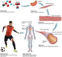
Additional info: The organization of the human body progresses from chemical, cellular, tissue, organ, organ system, to organismal levels.
Generalized Cell Structure
Basic Parts of a Human Cell
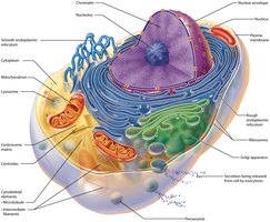
All human cells share three main components:
Plasma membrane: Flexible outer boundary that separates the cell from its environment.
Cytoplasm: Intracellular fluid containing organelles.
Nucleus: Control center containing DNA.
Extracellular Materials
Types and Functions
Extracellular materials are substances found outside cells, including:
Extracellular fluids: Interstitial fluid (bathes cells), blood plasma, cerebrospinal fluid.
Cellular secretions: Such as saliva and mucus.
Extracellular matrix (ECM): A network of proteins and polysaccharides that provides structural support and acts as a "glue" to hold cells together.
Plasma Membrane
Structure and Function
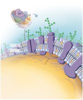
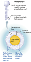
The plasma membrane is a dynamic barrier that separates intracellular fluid from extracellular fluid. It controls what enters and exits the cell and is described by the fluid mosaic model, where proteins float in a sea of lipids.
Physical barrier: Encloses the cell, maintaining internal environment.
Selective permeability: Regulates entry and exit of substances.
Communication: Membrane proteins interact with chemical messengers.
Cell recognition: Carbohydrates on the surface allow cells to identify each other.
Membrane Lipids
The lipid bilayer is primarily composed of phospholipids, with cholesterol and glycolipids interspersed.
Phospholipids: Have hydrophilic (water-loving) heads and hydrophobic (water-fearing) tails, forming a bilayer.
Cholesterol: Stabilizes membrane fluidity and structure.
Membrane Proteins
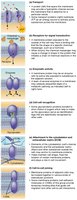
Membrane proteins are essential for communication and transport. They are classified as:
Integral proteins: Embedded in the membrane, often spanning it (transmembrane).
Peripheral proteins: Loosely attached to the membrane surface or to integral proteins.
Functions include transport, signal transduction, enzymatic activity, cell-cell recognition, attachment to cytoskeleton/ECM, and intercellular joining.
Glycocalyx
The glycocalyx is a carbohydrate-rich area on the cell surface, functioning as a biological marker for cell recognition and immune response. It is composed of glycoproteins and glycolipids.
Clinical Note: Cancer cells may alter their glycocalyx, evading immune detection.
Cell Junctions
Types and Functions
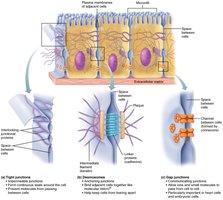
Cell junctions connect adjacent cells and facilitate communication:
Tight junctions: Form impermeable barriers (e.g., in intestines, bladder).
Desmosomes: Provide mechanical strength (e.g., skin, cardiac muscle).
Gap junctions: Allow passage of ions and small molecules for communication (e.g., heart, smooth muscle).
Membrane Transport
Overview
The plasma membrane is selectively permeable, allowing some substances to cross more easily than others. Transport occurs via passive or active mechanisms.
Passive transport: No energy required; includes simple diffusion, facilitated diffusion, and osmosis.
Active transport: Requires ATP; includes primary/secondary active transport and vesicular transport.
Passive Membrane Transport
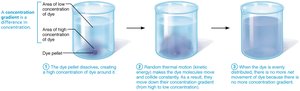
Passive transport relies on the movement of molecules down their concentration gradients.
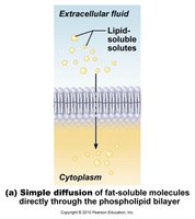
Simple diffusion: Movement of nonpolar, lipid-soluble molecules directly through the bilayer (e.g., O2, CO2).
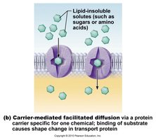
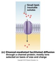
Facilitated diffusion: Movement of polar or charged molecules via carrier or channel proteins (e.g., glucose, ions).
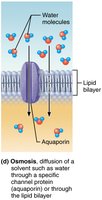
Osmosis: Diffusion of water through a selectively permeable membrane, often via aquaporins.
Osmolarity and Tonicity
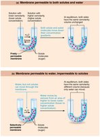
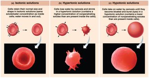
Osmolarity is the total concentration of solute particles in a solution. Tonicity describes how a solution affects cell volume:
Isotonic: No net water movement; cell volume unchanged.
Hypertonic: Water leaves cell; cell shrinks (crenation).
Hypotonic: Water enters cell; cell swells and may burst (lysis).
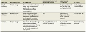
Passive Membrane Transport Table
Process | Energy Source | Description | Membrane Transport Protein Required | Specific and Saturable | Examples |
|---|---|---|---|---|---|
Simple diffusion | Kinetic energy | Net movement of molecules down concentration gradient | No | No | Lipids, oxygen, carbon dioxide |
Facilitated diffusion | Kinetic energy | Diffusing substance attached to membrane carrier or moves through channel | Yes | Yes | Glucose, Na+, K+ |
Osmosis | Kinetic energy | Diffusion of water through a selectively permeable membrane | No (except aquaporins) | No | Water |
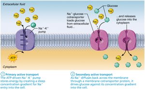
Active Membrane Transport
Active transport requires energy (ATP) to move substances against their concentration gradients or to transport large substances.
Primary active transport: Direct use of ATP (e.g., sodium-potassium pump).
Secondary active transport: Indirect use of ATP via ion gradients.
Vesicular transport: Movement of large particles or fluids via vesicles (endocytosis, exocytosis, transcytosis).
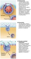
Vesicular Transport
Vesicular transport moves large substances or large volumes across membranes using vesicles and ATP.
Endocytosis: Import into cell (phagocytosis, pinocytosis, receptor-mediated endocytosis).
Exocytosis: Export from cell (e.g., secretion of hormones, neurotransmitters).
Transcytosis: Movement into, across, and out of the cell.
Vesicular trafficking: Movement within the cell.
Membrane Potential
Resting Membrane Potential (RMP)
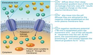
The resting membrane potential is the voltage difference across the plasma membrane, typically –50 to –90 mV, maintained by sodium-potassium pumps. It is essential for nerve and muscle function.
Cytoplasm and Organelles
Cytoplasmic Components
The cytoplasm contains cytosol (fluid), inclusions (stored substances), and organelles (specialized structures).
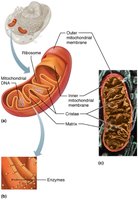
Mitochondria: Site of ATP production via aerobic respiration.
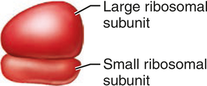
Ribosomes: Sites of protein synthesis; free or membrane-bound.
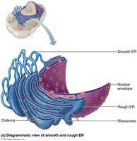
Endoplasmic reticulum (ER): Rough ER synthesizes proteins; smooth ER synthesizes lipids and detoxifies chemicals.
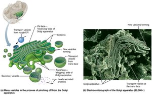
Golgi apparatus: Modifies, sorts, and packages proteins and lipids.
Peroxisomes: Detoxify harmful substances.
Lysosomes: Digest cellular debris and foreign material.
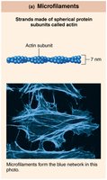
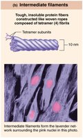
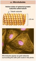
Cytoskeleton: Provides structural support and facilitates movement (microfilaments, intermediate filaments, microtubules).
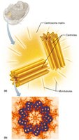
Centrosome and centrioles: Organize microtubules and cell division.
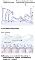
Cilia and flagella: Motile extensions for movement.
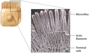
Microvilli: Increase surface area for absorption.
Nucleus
Structure and Function
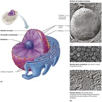
The nucleus is the largest organelle, containing the genetic material (DNA) and controlling cellular activities. Most cells are uninucleate, but some are multinucleate or anucleate.
Nuclear envelope: Double membrane with pores for molecular exchange.
Nucleolus: Site of ribosomal RNA synthesis and ribosome assembly.
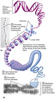
Chromatin: DNA-protein complex; condenses to form chromosomes during cell division.
Cell Cycle
Phases and Control
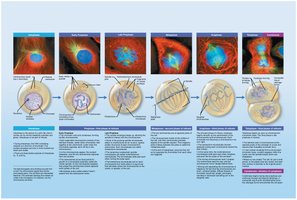
The cell cycle describes the life of a cell from formation to division. It consists of interphase (growth and DNA replication) and the mitotic phase (mitosis and cytokinesis).
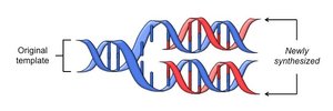
Interphase: G1 (growth), S (DNA synthesis), G2 (preparation for division).
Mitosis: Prophase, metaphase, anaphase, telophase.
Cytokinesis: Division of cytoplasm.
Checkpoints: Ensure proper division; G1 checkpoint is most critical.
Protein Synthesis
DNA, RNA, and the Central Dogma
Protein synthesis is directed by DNA through two main processes:
Transcription: DNA is copied into messenger RNA (mRNA) in the nucleus.
Translation: mRNA is decoded by ribosomes in the cytoplasm to assemble amino acids into proteins.
Types of RNA involved:
mRNA: Carries genetic code from DNA to ribosomes.
rRNA: Structural component of ribosomes.
tRNA: Brings amino acids to ribosomes during translation.
Clinical Note: mRNA vaccines use synthetic mRNA to instruct cells to produce viral proteins, stimulating an immune response.
Cellular Maintenance and Death
Autophagy, Proteasomes, and Apoptosis
Cells maintain homeostasis by recycling or destroying damaged components:
Autophagy: Lysosomal degradation of nonfunctional organelles.
Proteasomes: Degrade ubiquitin-tagged proteins.
Apoptosis: Programmed cell death, essential for development and disease prevention.
Developmental Aspects and Cell Growth
Cell Differentiation and Growth
All cells contain the same DNA but express different genes, leading to structural and functional diversity (cell differentiation). Cell growth can occur by increasing cell number (hyperplasia) or cell size (hypertrophy). Atrophy is a decrease in cell size, and necrosis is uncontrolled cell death due to injury or disease.
