 뒤로
뒤로Cells: The Living Units – Structure, Function, and Membrane Transport
스터디 가이드 - 스마트 노트
 자료에 맞춘 맞춤형 노트, 핵심 정의, 예시, 맥락을 확장해 제공합니다.
3.1 Cells: The Smallest Living Units
Cell Theory
Cell theory is a foundational concept in biology, emphasizing that the cell is the basic structural and functional unit of life. The activities of an organism depend on the individual and collective activities of its cells. The structure of each cell is closely related to its function, and all cells arise from preexisting cells, ensuring the continuity of life.
Structural and Functional Unit: All living things are composed of cells, which perform essential life processes.
Complementarity of Structure and Function: The shape and internal structures of a cell determine its biochemical activities.
Cellular Continuity: New cells are produced only by the division of existing cells.
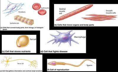
Cell Diversity
Human bodies contain over 250 different types of cells, each specialized for particular functions. These differences in size, shape, and subcellular components enable the wide variety of physiological roles necessary for life.
Examples: Muscle cells (movement), nerve cells (information transmission), fat cells (nutrient storage), macrophages (defense), sperm cells (reproduction).
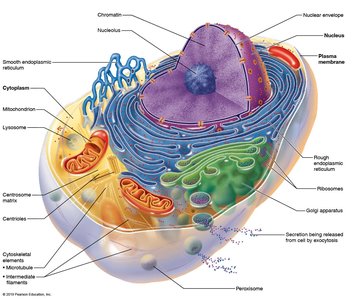
Structure of the Generalized Cell
Despite their diversity, all human cells share three basic structural components:
Plasma membrane: The flexible outer boundary that separates the cell from its environment.
Cytoplasm: The intracellular fluid containing organelles.
Nucleus: The control center containing DNA.
Extracellular Materials
Substances found outside cells are collectively known as extracellular materials. These include:
Extracellular fluids: Interstitial fluid (bathes cells), blood plasma (in blood), cerebrospinal fluid (around nervous tissue).
Cellular secretions: Such as saliva and mucus.
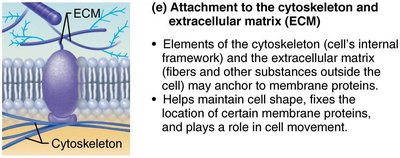
Extracellular matrix: A network of proteins and polysaccharides that provides structural support and acts as a "glue" to hold cells together.
3.2 Structure of the Plasma Membrane
The Plasma Membrane
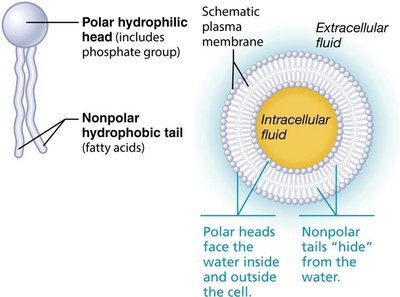
The plasma membrane, also known as the cell membrane, is a dynamic barrier that separates the intracellular fluid (ICF) from the extracellular fluid (ECF). It controls what enters and exits the cell, maintaining homeostasis.
Fluid Mosaic Model: The membrane is composed of a flexible lipid bilayer with proteins floating within it, creating a constantly changing mosaic pattern.
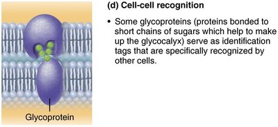
Glycocalyx: Surface sugars that play roles in cell recognition and adhesion.
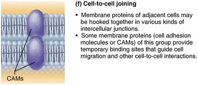
Cell Junctions: Structures that connect adjacent cells.
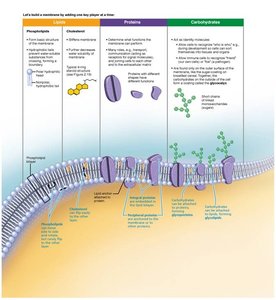
Membrane Lipids
The lipid bilayer forms the basic structure of the plasma membrane:
Phospholipids (75%): Have hydrophilic (water-loving) heads and hydrophobic (water-fearing) tails, forming a bilayer.
Glycolipids (5%): Lipids with attached sugars, found on the outer surface.
Cholesterol (20%): Stabilizes the membrane and maintains its fluidity.
Membrane Proteins
Proteins make up about half the mass of the plasma membrane and are responsible for most of its specialized functions. They can be:
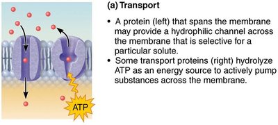
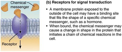
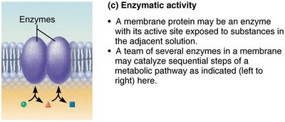
Integral proteins: Firmly embedded in the membrane, often spanning it (transmembrane). Functions include transport, acting as enzymes, or receptors.
Peripheral proteins: Loosely attached to the membrane surface. Functions include acting as enzymes, motor proteins, or in cell-to-cell connections.
Cell Junctions
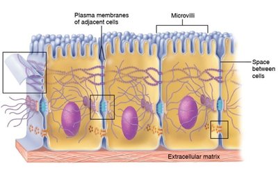
Most cells are bound together to form tissues and organs. There are three main types of cell junctions:
Tight junctions: Seal adjacent cells to prevent passage of molecules between them.
Desmosomes: Anchor cells together, providing mechanical stability.
Gap junctions: Allow communication and passage of ions and small molecules between cells.
3.3 Passive Membrane Transport
Overview of Passive Transport
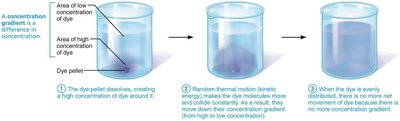
Passive transport does not require cellular energy (ATP). It relies on the natural movement of molecules from areas of high concentration to low concentration, a process known as diffusion.
Diffusion: Driven by the intrinsic kinetic energy of molecules, resulting in their movement down a concentration gradient.
Simple Diffusion
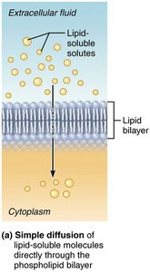
Simple diffusion involves the movement of nonpolar, lipid-soluble substances directly through the phospholipid bilayer. Examples include oxygen, carbon dioxide, steroid hormones, and fatty acids. Small amounts of very small polar substances, such as water, can also pass through.
Facilitated Diffusion
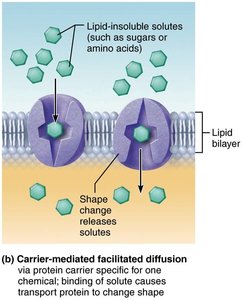
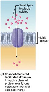
Facilitated diffusion is used for molecules that are not lipid-soluble or are too large to pass through the membrane pores. It occurs via:
Carrier-mediated facilitated diffusion: Substances bind to protein carriers, which change shape to transport the molecules across the membrane.
Channel-mediated facilitated diffusion: Substances move through water-filled protein channels. Channels can be always open (leaky) or gated (regulated by signals).
Osmosis
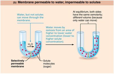
Osmosis is the diffusion of a solvent, such as water, across a selectively permeable membrane. Water moves through the lipid bilayer or via specific channels called aquaporins. The direction of water movement depends on the relative concentrations of solutes on either side of the membrane.
Osmolarity: The total concentration of solute particles in a solution.
Water moves from areas of low solute concentration (high water) to high solute concentration (low water).
Influence of Membrane Permeability on Diffusion and Osmosis
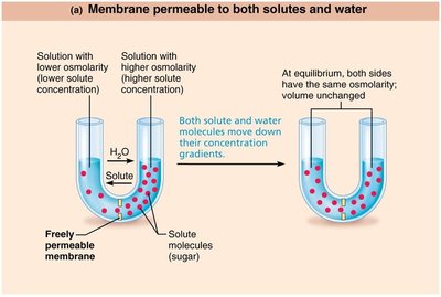
The permeability of the membrane determines whether solutes, water, or both can move across. If only water can move, osmosis will occur until equilibrium is reached, resulting in volume changes.
Tonicity
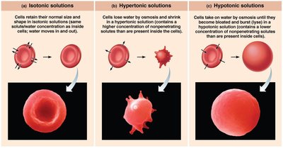
Tonicity describes how a solution affects cell volume:
Isotonic: Same osmolarity as the cell; no net water movement.
Hypertonic: Higher osmolarity than the cell; water leaves the cell, causing it to shrink.
Hypotonic: Lower osmolarity than the cell; water enters the cell, causing it to swell.
3.4 Active Membrane Transport
Overview of Active Transport
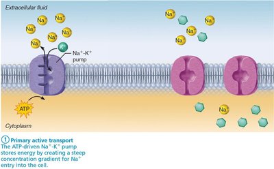
Active transport requires energy (usually ATP) to move solutes against their concentration gradients (from low to high concentration). This process is essential for maintaining cellular homeostasis.
Primary active transport: Direct use of ATP to transport molecules (e.g., Na+-K+ pump).
Secondary active transport: Indirect use of ATP, relying on gradients established by primary active transport.
Vesicular Transport
Vesicular transport moves large particles, macromolecules, and fluids across the membrane in vesicles. This process requires energy and includes:
Endocytosis: Transport into the cell (phagocytosis, pinocytosis, receptor-mediated endocytosis).
Exocytosis: Ejection of substances from the cell.
Transcytosis: Movement into, across, and out of the cell.
Vesicular trafficking: Movement of substances within the cell.
3.5 Membrane Potential
Resting Membrane Potential (RMP)
The resting membrane potential is the electrical potential energy resulting from the separation of oppositely charged particles across the plasma membrane. In most cells, the inside is more negative relative to the outside, with voltages ranging from –50 to –100 mV. The electrochemical gradient of potassium (K+) is the primary determinant of RMP.
3.6 Cell-Environment Interactions
Cell Adhesion Molecules (CAMs) and Plasma Membrane Receptors
Cells interact with their environment through direct contact with other cells or by responding to extracellular chemicals. Key mechanisms include:
Cell adhesion molecules (CAMs): Glycoproteins that anchor cells to the extracellular matrix or to each other, and attract white blood cells to sites of injury or infection.
Plasma membrane receptors: Proteins that serve as binding sites for chemical signals (ligands) such as neurotransmitters, hormones, and paracrines. Ligand-receptor interactions trigger changes in cellular activity.
