 뒤로
뒤로Cellular Level of Organization: Structure and Function in Anatomy & Physiology
스터디 가이드 - 스마트 노트
 자료에 맞춘 맞춤형 노트, 핵심 정의, 예시, 맥락을 확장해 제공합니다.
Fundamentals of Anatomy & Physiology
Introduction to Cells
Cells are the fundamental units of life in the human body, forming the basis for all physiological functions. Understanding cell structure and function is essential for grasping the principles of anatomy and physiology.
Smallest Living Units: Cells are eukaryotic and are the smallest units capable of life.
Building Blocks: All organisms are composed of cells.
Cell Division: New cells arise from the division of preexisting cells.
Homeostasis: Each cell maintains its own internal balance, contributing to overall organismal homeostasis.
Trillions of Cells: The human body contains trillions of cells, including numerous microbial cells.
Plasma Membrane
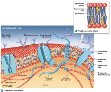
The plasma membrane is a selectively permeable barrier that separates the cell's internal environment from the external environment. It is crucial for maintaining cellular integrity and regulating transport.
Structure: Composed mainly of a phospholipid bilayer and cholesterol.
Functions: Physical separation, regulation of transport, sensitivity (cell communication), and support (anchoring cells).
Phospholipid Bilayer: Hydrophilic heads face outward toward water, hydrophobic tails face inward, creating a barrier to water-soluble substances.
Cholesterol: Adds rigidity and reduces permeability.
Proteins: Integral (embedded), transmembrane (span the membrane), and peripheral (surface-bound) proteins perform various functions.
Eukaryotic Cell Organelles
Organelles are specialized structures within cells that perform distinct functions. They are classified as nonmembranous or membranous based on their structure.
Cytoplasm: Includes cytosol (intracellular fluid), organelles, and inclusions (insoluble masses like glycogen, lipid droplets, and pigment granules).
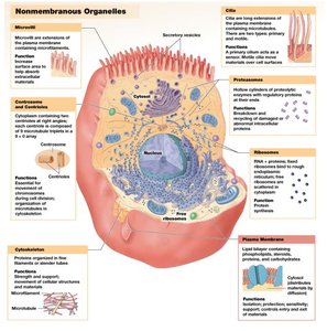
Nonmembranous Organelles: Not enclosed by membranes; include cytoskeleton, centrioles, and ribosomes.
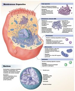
Membranous Organelles: Enclosed by phospholipid membranes; include endoplasmic reticulum (ER), Golgi apparatus, lysosomes, peroxisomes, nucleus, and mitochondria.
Cytoskeleton
The cytoskeleton is an internal network of protein fibers that provides structural support, facilitates movement, and organizes cell contents.
Microfilaments: Smallest fibers, composed of actin; provide mechanical strength and are involved in muscle contraction.
Intermediate Filaments: Provide strength, maintain cell shape, and stabilize organelle positions.
Microtubules: Largest fibers, hollow tubes of tubulin; radiate from the centrosome, anchor organelles, and form spindle apparatus during cell division.
Ribosomes
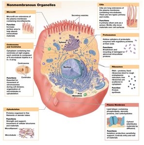
Ribosomes are the sites of protein synthesis. They can be free in the cytoplasm or fixed to the rough ER.
Structure: Composed of small and large subunits containing rRNA and proteins.
Free Ribosomes: Synthesize proteins for use in the cytosol.
Fixed Ribosomes: Attached to the rough ER; synthesize proteins for modification and export.
Endoplasmic Reticulum (ER)
The ER is a network of membranes involved in synthesis, storage, transport, and detoxification.
Smooth ER (SER): Lacks ribosomes; synthesizes phospholipids, cholesterol, steroid hormones, triglycerides, glycogen, stores Ca2+, and detoxifies drugs.
Rough ER (RER): Has ribosomes; synthesizes and folds proteins, packages them into vesicles for the Golgi apparatus.
Golgi Apparatus
The Golgi apparatus modifies, packages, and sorts proteins and lipids for secretion or use within the cell.
Structure: Series of flattened membranous discs (cisternae).
Functions: Modifies proteins, packages secretions, forms lysosomes.
Lysosomes
Lysosomes are membrane-bound vesicles containing digestive enzymes. They break down cellular debris, damaged organelles, and foreign substances.
Functions: Hydrolyze polymers, recycle organelles, destroy bacteria, and perform autolysis (self-destruction).
Mitochondria
Mitochondria are the powerhouses of the cell, producing ATP through cellular respiration.
Structure: Double-membraned organelle.
Function: Energy production; number varies by cell type based on energy demand.
Nucleus
The nucleus is the control center of the cell, storing genetic information and regulating protein synthesis.
Nuclear Envelope: Double membrane with nuclear pores for communication with cytoplasm.
Nucleolus: Synthesizes rRNA and assembles ribosomal subunits.
Chromatin: Loosely coiled DNA in non-dividing cells; chromosomes in dividing cells.
Protein Synthesis
Protein synthesis follows the central dogma: DNA is transcribed to mRNA, which is then translated into protein.
Transcription: DNA → mRNA in the nucleus.
RNA Processing: Introns removed, exons spliced; alternative splicing allows multiple proteins from one gene.
Translation: mRNA → polypeptide at ribosomes; tRNA delivers amino acids based on codon-anticodon pairing.
Transport Across the Plasma Membrane
Cells regulate the movement of substances across the plasma membrane using passive and active mechanisms.
Passive Transport: No energy required; includes diffusion and osmosis.
Active Transport: Requires energy (ATP); includes ion pumps and sodium-potassium exchange pump.
Carrier-Mediated Transport: Uses specialized proteins; can be passive (facilitated diffusion) or active.
Symporters: Move two substances in the same direction.
Antiporters: Move two substances in opposite directions.
Diffusion and Osmosis
Diffusion is the movement of substances from high to low concentration. Osmosis is the diffusion of water across a selectively permeable membrane.
Simple Diffusion: Lipid-soluble substances pass directly through the membrane.
Channel-Mediated Diffusion: Water-soluble substances pass through protein channels.
Osmosis: Water moves toward higher solute concentration.
Tonicity
Tonicity describes how solute concentration affects cell size and shape.
Isotonic: Equal solute concentrations; cell size remains unchanged.
Hypotonic: Lower solute concentration outside; cell swells and may burst (hemolysis).
Hypertonic: Higher solute concentration outside; cell shrivels (crenation).
Cell Life Cycle
Cell division is essential for growth, repair, and maintenance. The cell cycle includes interphase (G1, S, G2), mitosis, and cytokinesis.
Interphase: Cell grows, duplicates organelles, and replicates DNA.
G0 Phase: Nondividing state; some cells remain here indefinitely.
Mitosis: Nuclear division; stages include prophase, metaphase, anaphase, and telophase.
Cytokinesis: Division of cytoplasm; cleavage furrow forms and separates cells.
Stage | Main Event |
|---|---|
Prophase | Chromosomes condense, nuclear envelope disintegrates, spindle fibers form |
Metaphase | Chromosomes align at metaphase plate |
Anaphase | Sister chromatids separate, move to opposite poles |
Telophase | Nuclear membranes reform, chromosomes decondense |
Cytokinesis | Cytoplasm divides, two daughter cells form |
Example: In human skin, cells constantly divide to replace those lost to abrasion, demonstrating the importance of the cell cycle in tissue maintenance.
