 뒤로
뒤로Cellular Structure and Membrane Transport: Study Notes for Anatomy & Physiology
스터디 가이드 - 스마트 노트
 자료에 맞춘 맞춤형 노트, 핵심 정의, 예시, 맥락을 확장해 제공합니다.
Cell Junctions
Types of Cell Junctions
Cell junctions are specialized structures that connect cells to one another, forming tissues and organs. Most cells are bound together by these junctions, which serve various structural and functional purposes.
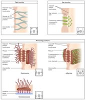
Tight Junctions: Integral proteins fuse adjacent cells, forming an impermeable barrier that prevents fluids and most molecules from passing between cells. This compartmentalizes body regions and is especially important in epithelial tissues, such as the lining of the intestines.
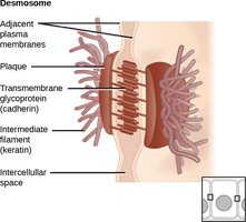
Desmosomes: Rivet-like junctions formed by cadherin proteins interlocking between cells. These are anchored to plaques inside the plasma membrane and connected by keratin filaments, providing mechanical strength and allowing flexibility to prevent tearing under tension. Common in tissues subject to mechanical stress, such as skin and heart muscle.
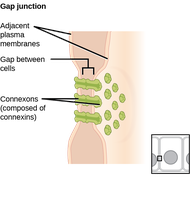
Gap Junctions: Connexon proteins form channels that allow ions and small molecules to pass directly between cells. These junctions facilitate rapid communication, especially in cardiac and smooth muscle cells.
Tight Junctions
Tight junctions create a seal around cells, preventing leakage of extracellular fluid and maintaining distinct compartments within tissues.
Function: Prevents passage of substances between cells.
Location: Found in epithelial tissues lining organs and cavities.
Desmosomes
Desmosomes provide strong adhesion between cells, allowing tissues to withstand mechanical stress.
Structure: Linker proteins (cadherins) interlock, anchored to plaques and connected by keratin filaments.
Function: Allows flexibility and prevents tearing.
Location: Skin, heart muscle.
Gap Junctions
Gap junctions enable direct communication between cells by allowing ions and small molecules to pass through connexon channels.
Function: Facilitates electrical and chemical signaling.
Location: Cardiac and smooth muscle cells.
Structure of the Plasma Membrane
Selective Permeability
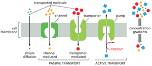
The plasma membrane is a selectively permeable barrier, allowing only certain molecules to cross. Substances move across the membrane via passive or active transport.
Passive Transport: No energy required; substances move down their concentration gradient.
Active Transport: Requires energy (ATP); substances move against their concentration gradient.
Passive Processes
Diffusion
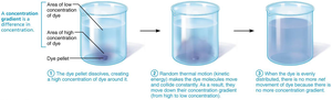
Diffusion is the movement of molecules from areas of high concentration to low concentration due to kinetic energy. Equilibrium is reached when there is no net movement.
Factors Influencing Diffusion: Concentration gradient, molecular size, temperature.
Types: Simple diffusion (directly through lipid bilayer), facilitated diffusion (via carrier or channel proteins), osmosis (movement of water).
Channel-Mediated Diffusion
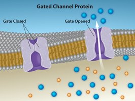
Transmembrane proteins form channels for ions or water to pass down their concentration gradient. Channels may be always open (leakage) or gated (controlled by signals).
Specificity: Based on pore size and charge.
Water Channels: Called aquaporins.
Carrier-Mediated Diffusion
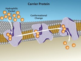
Carrier proteins transport specific polar molecules (e.g., sugars, amino acids) across the membrane by changing shape upon binding.
Saturation: Limited by number of carriers; all carriers can be occupied.
Osmosis
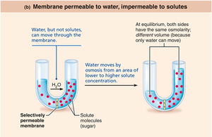
Osmosis is the movement of water across a selectively permeable membrane from areas of low solute concentration to high solute concentration.
Osmolarity: Measure of solute concentration.
Direction: Water moves to equalize solute concentrations.
Tonicity
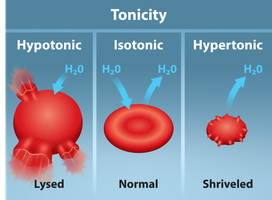
Tonicity describes the effect of a solution on cell shape by altering internal water volume.
Isotonic: Same osmolarity as cell; no change in volume.
Hypertonic: Higher osmolarity; cell shrinks (crenation).
Hypotonic: Lower osmolarity; cell swells (lysis).
Active Membrane Transport
Active Transport
Active transport uses carrier proteins (solute pumps) to move substances against their concentration gradient, requiring ATP.
Antiporters: Transport one substance in and another out.
Symporters: Transport two substances in the same direction.
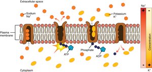
Primary Active Transport: Direct use of ATP (e.g., Na+-K+ pump).
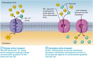
Secondary Active Transport: Indirect use of ATP via ion gradients.
Primary and Secondary Active Transport
The sodium-potassium pump (Na+-K+ ATPase) maintains electrochemical gradients by pumping 3 Na+ out and 2 K+ in per ATP molecule. Secondary active transport uses the gradient established by primary transport to move other molecules (e.g., Na+-glucose symporter).
Electrochemical Gradient: Essential for muscle and nerve function.
Example: Na+-glucose cotransporter.
Vesicular Transport
Types of Vesicular Transport
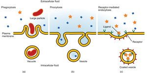
Vesicular transport moves large particles, macromolecules, and fluids across membranes in vesicles, requiring ATP.
Endocytosis: Transport into cell (phagocytosis, pinocytosis, receptor-mediated).
Exocytosis: Transport out of cell.
Transcytosis: Transport across cell.
Vesicular Trafficking: Transport within cell.
Membrane Potential
Resting Membrane Potential (RMP)
RMP is the electrical potential across the plasma membrane in resting cells, typically –50 to –100 mV. It is established by the movement of ions, mainly K+, and maintained by the Na+-K+ pump.
Polarization: Inside of cell is more negative than outside.
Electrochemical Gradient: Drives ion movement.
Cytoplasm and Organelles
Cytoplasm
The cytoplasm is the material between the plasma membrane and nucleus, containing cytosol, inclusions, and organelles.
Cytosol: Gel-like solution with water, proteins, salts, sugars.
Inclusions: Insoluble molecules (e.g., glycogen, pigments).
Organelles: Metabolic machinery, either membranous or nonmembranous.
Membranous Organelles
Mitochondria: ATP production via aerobic respiration; double membrane; contains own DNA.
Endoplasmic Reticulum (ER): Rough ER (protein synthesis), Smooth ER (lipid synthesis, detoxification).
Golgi Apparatus: Modifies, packages, and secretes proteins and lipids.
Peroxisomes: Detoxify substances; break down fatty acids.
Lysosomes: Digestive enzymes; degrade organelles and pathogens.
Nonmembranous Organelles
Ribosomes: Protein synthesis; free or membrane-bound.
Cytoskeleton: Microfilaments, intermediate filaments, microtubules; structural support and movement.
Centrioles: Cell division; basis of cilia and flagella.
Cytoskeleton
Types of Cytoskeletal Elements
Microfilaments: Actin; cell shape, movement.
Intermediate Filaments: Ropelike; mechanical strength.
Microtubules: Tubulin; organelle distribution, tracks for motor proteins.
Cellular Extensions
Cilia, Flagella, and Microvilli
Cilia and flagella are microtubule-based extensions for movement; microvilli are actin-based projections for absorption.
Cilia: Move substances across cell surface.
Flagella: Propel entire cell (e.g., sperm).
Microvilli: Increase surface area for absorption.
Nucleus
Structure and Function
The nucleus is the largest organelle, containing genetic material for protein synthesis. It has a double-membrane envelope, nucleoli for rRNA synthesis, and chromatin for DNA packaging.
Nuclear Envelope: Double membrane with pores for transport.
Nucleolus: Ribosome assembly.
Chromatin: DNA and histone proteins; condenses to form chromosomes during cell division.
Discussion Questions
Impact of Drinking Seawater: Seawater is hypertonic compared to body fluids, causing cells to lose water and shrink (crenation).
Lysosomes vs. Peroxisomes: Lysosomes digest cellular debris and pathogens; peroxisomes detoxify substances and break down fatty acids.
Compare Cilia, Flagella, Microvilli: Cilia move substances, flagella move cells, microvilli increase absorption surface area.
