 뒤로
뒤로Chapter 16: The Endocrine System – Anatomy & Physiology Study Notes
스터디 가이드 - 스마트 노트
 자료에 맞춘 맞춤형 노트, 핵심 정의, 예시, 맥락을 확장해 제공합니다.
Overview of the Endocrine System
Introduction
The endocrine system is a complex network of organs that synthesize and secrete chemical messengers called hormones. These hormones interact with specific target cells to regulate vital functions such as fluid, electrolyte, and acid-base homeostasis, growth, and metabolic reactions.
Hormones: Chemical messengers secreted into the blood to affect distant targets.
Paracrine signals: Chemicals secreted by cells into extracellular space, affecting nearby but different cell types.
Autocrine signals: Chemicals secreted by cells into interstitial fluid, eliciting effects from the same cell or cell type.
Endocrine Organs
Endocrine glands are ductless organs that regulate other cell types by producing and secreting hormones.
Primary endocrine organs: Anterior pituitary, thyroid, parathyroid, adrenal cortices, endocrine pancreas, thymus.
Secondary endocrine organs: Heart, kidneys, small intestines, testes, ovaries.
Neuroendocrine organs: Hypothalamus, pineal gland, adrenal medulla.
Hormones: Structure, Transport, and Action
Classes of Hormones
Hormones are classified by their chemical structure:
Amino-acid hormones: Hydrophilic, bind to plasma membrane receptors.
Steroid hormones: Derived from cholesterol, hydrophobic, bind to receptors in cytosol or nucleus.
Hormone Transport
Free hormones: Small, hydrophilic, travel freely in blood plasma.
Bound hormones: Hydrophobic, form complexes with plasma proteins, extend hormone lifespan and regulate concentration.
Target Cells and Receptors
Hormones affect only target cells with specific protein receptors.
Hydrophilic hormones: Bind to membrane receptors.
Hydrophobic hormones: Cross plasma membrane, bind to intracellular receptors.
Up-regulation: Increased receptor number when hormone levels decline.
Down-regulation: Decreased receptor number with prolonged high hormone levels.
Mechanisms of Hormone Action
Hydrophilic hormones: Bind to membrane receptors, activate G-proteins, trigger second messengers (e.g., cAMP), activate protein kinases.
Hydrophobic hormones: Cross membrane, bind to intracellular receptors, influence gene transcription and protein synthesis.
The Hypothalamus and Pituitary Gland
Anatomy and Functional Relationship
The hypothalamus regulates many homeostatic processes and is connected to the pituitary gland by the infundibulum. The pituitary gland consists of:
Anterior pituitary (adenohypophysis): Glandular epithelium, secretes hormones.
Posterior pituitary (neurohypophysis): Nervous tissue, stores and releases neurohormones produced by the hypothalamus.
Hypothalamic-Hypophyseal Portal System
Specialized blood supply allows direct hormone delivery between hypothalamus and pituitary.
Hormones of the Hypothalamus and Pituitary Gland
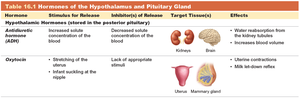
Posterior pituitary: Stores and releases antidiuretic hormone (ADH) and oxytocin.
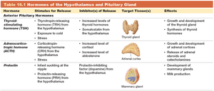
Anterior pituitary: Secretes tropic hormones that regulate other endocrine glands.
Hormone | Stimulus for Release | Inhibitor(s) of Release | Target Tissue(s) | Effects |
|---|---|---|---|---|
Antidiuretic Hormone (ADH) | Increased solute concentration of blood | Decreased solute concentration of blood | Kidneys, Brain | Water reabsorption, increases blood volume |
Oxytocin | Stretching of uterus, infant suckling | Lack of appropriate stimuli | Uterus, Mammary gland | Uterine contractions, milk let-down reflex |
Anterior Pituitary Hormones
Hormone | Stimulus for Release | Inhibitor(s) of Release | Target Tissue(s) | Effects |
|---|---|---|---|---|
Thyroid-stimulating hormone (TSH) | Thyrotropin-releasing hormone (TRH) | Increased levels of thyroid hormones, somatostatin | Thyroid gland | Growth and development of thyroid, synthesis of thyroid hormones |
Adrenocorticotropic hormone (ACTH) | Corticotropin-releasing hormone (CRH) | Increased level of cortisol | Adrenal cortex | Growth and development of adrenal cortices, release of adrenal steroids and catecholamines |
Prolactin | Infant suckling at nipple | Prolactin-inhibiting factor (dopamine) | Mammary gland | Development of mammary glands, milk production |
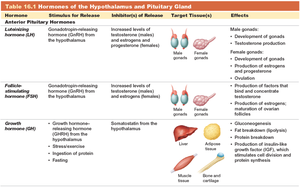
Hormone | Stimulus for Release | Inhibitor(s) of Release | Target Tissue(s) | Effects |
|---|---|---|---|---|
Luteinizing hormone (LH) | Gonadotropin-releasing hormone (GnRH) | Increased levels of testosterone (males) and estrogen/progesterone (females) | Male and female gonads | Development of gonads, testosterone production, production of estrogen and progesterone |
Follicle-stimulating hormone (FSH) | Gonadotropin-releasing hormone (GnRH) | Increased levels of testosterone and estrogen | Male and female gonads | Production of factors that bind and concentrate testosterone, production of estrogen, maturation of ovarian follicles |
Growth hormone (GH) | Growth hormone-releasing hormone (GHRH), stress, exercise, protein intake, fasting | Somatostatin from hypothalamus | Liver, adipose tissue, muscle, bone, cartilage | Growth, fat breakdown, production of IGF, protein synthesis |
The Thyroid and Parathyroid Glands
Structure and Function
Thyroid gland: Located in anterior neck, secretes thyroid hormone and calcitonin.
Parathyroid glands: Located on posterior surface of thyroid, secrete parathyroid hormone (PTH).
Thyroid Hormone
Consists of amino acid core bound to iodine atoms (T3 and T4).
Regulates metabolic rate, growth, and development.
Production regulated by a three-tiered negative feedback loop: TRH (hypothalamus) → TSH (anterior pituitary) → thyroid hormone (thyroid gland).
Thyroid Disorders
Hyperthyroidism: Overproduction, causes weight loss, heat intolerance, goiter, exophthalmos.
Hypothyroidism: Underproduction, causes weight gain, cold intolerance, slow heart rate, goiter.
Parathyroid Hormone and Calcitonin
PTH: Increases blood calcium by stimulating osteoclasts, increasing intestinal absorption, and kidney reabsorption.
Calcitonin: Lowers blood calcium by inhibiting osteoclast activity.
The Adrenal Glands
Structure
Adrenal glands are located on the superior aspect of each kidney and consist of an outer cortex and inner medulla.
Cortex: Produces steroid hormones (mineralocorticoids, glucocorticoids, androgenic steroids).
Medulla: Secretes catecholamines (epinephrine, norepinephrine).
Hormones of the Adrenal Cortex
Cortisol: Regulates stress response, increases blood glucose, induces protein and fat breakdown, anti-inflammatory.
Regulation involves the HPA axis: CRH (hypothalamus) → ACTH (anterior pituitary) → cortisol (adrenal cortex).
Cortisol Disorders
Cushing’s disease: Oversecretion, causes fat redistribution, muscle wasting, hypertension, immunosuppression, osteoporosis.
The Endocrine Pancreas
Structure
The pancreas is both an endocrine and exocrine gland.
Pancreatic islets: Contain alpha (glucagon), beta (insulin), and delta (somatostatin) cells.
Hormones and Regulation
Glucagon: Increases blood glucose by promoting glycogenolysis, gluconeogenesis, and fat breakdown.
Insulin: Lowers blood glucose by promoting uptake and storage of nutrients.
Feedback loops maintain blood glucose homeostasis.
Diabetes Mellitus
Type 1: Destruction of beta cells, insulin deficiency, risk of ketoacidosis.
Type 2: Insulin resistance, associated with obesity and heredity.
Chronic hyperglycemia damages blood vessels, nerves, eyes, and kidneys.
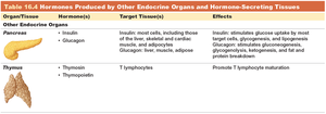
Organ | Hormone(s) | Target Tissue(s) | Effects |
|---|---|---|---|
Pancreas | Insulin, Glucagon | Most cells, liver, muscle, adipose | Insulin: glucose uptake, storage; Glucagon: glycogenolysis, gluconeogenesis, fat/protein breakdown |
Thymus | Thymosin, Thymopoietin | T lymphocytes | Promote T lymphocyte maturation |
Other Endocrine Glands and Hormone-Secreting Tissues
The Thymus
Secretes thymosin and thymopoietin, which assist in T lymphocyte maturation.
The Gonads
Testes: Produce testosterone, regulated by GnRH, LH, and FSH.
Ovaries: Produce estrogen and progesterone, regulated by multi-tiered feedback loops.
Sex hormones regulate gamete production, secondary sex characteristics, and reproductive functions.
Examples of Endocrine Control
Fluid Homeostasis
ADH, aldosterone, and ANP regulate water balance and urine output.
Negative feedback loops adjust hormone secretion based on plasma volume and solute concentration.
Metabolic Homeostasis
Thyroid hormones, glucagon, GH, insulin, and leptin regulate metabolism during fasting, feeding, and exercise.
Stress Response
Hormonal responses to stress involve cortisol and catecholamines, increasing metabolic rate and mobilizing energy stores.
Summary Table: Hormones of the Endocrine System
Hormone | Source | Target | Main Effect |
|---|---|---|---|
ADH | Posterior pituitary | Kidneys, brain | Water reabsorption, increased blood volume |
Oxytocin | Posterior pituitary | Uterus, mammary gland | Uterine contractions, milk let-down |
TSH | Anterior pituitary | Thyroid gland | Thyroid hormone synthesis |
ACTH | Anterior pituitary | Adrenal cortex | Adrenal steroid release |
Insulin | Pancreas | Most cells | Glucose uptake, storage |
Glucagon | Pancreas | Liver, muscle, adipose | Increase blood glucose |
Thymosin | Thymus | T lymphocytes | T cell maturation |
Key Equations
Gluconeogenesis:
Glycogenolysis:
Additional info:
Some details about hormone regulation and feedback loops were expanded for clarity.
Tables were recreated and summarized based on the original images and content.
