 뒤로
뒤로Chapter 17: Blood – Structure, Function, and Clinical Relevance
스터디 가이드 - 스마트 노트
 자료에 맞춘 맞춤형 노트, 핵심 정의, 예시, 맥락을 확장해 제공합니다.
Blood: Overview and Functions
Introduction to Blood
Blood is a specialized connective tissue that serves as the body's internal transport system. It is essential for maintaining homeostasis by transporting substances, regulating physiological parameters, and providing protection against disease.
Transport: Delivers oxygen and nutrients to cells, removes metabolic wastes, and transports hormones.
Regulation: Maintains body temperature, pH balance, and fluid volume.
Protection: Prevents blood loss via clotting and combats infection through immune cells and proteins.
Composition of Blood
Major Components
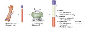
Blood consists of two main components: plasma (the liquid matrix) and formed elements (cells and cell fragments).
Plasma: Straw-colored fluid, mostly water, containing proteins, nutrients, hormones, and waste products.
Formed Elements: Includes erythrocytes (red blood cells), leukocytes (white blood cells), and platelets (cell fragments).
Physical Characteristics and Volume
Blood is more viscous than water and has a metallic taste.
Color varies with oxygen content: bright red (oxygen-rich), dark red (oxygen-poor).
pH: 7.35–7.45; volume: 5–6 L in males, 4–5 L in females.
Blood Plasma
Plasma is about 90% water and contains over 100 dissolved solutes, including:
Plasma proteins: Albumin (major contributor to osmotic pressure), globulins, fibrinogen.
Nutrients, gases, hormones, wastes, and electrolytes.
Formed Elements of Blood
Types and Features
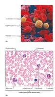
The formed elements include erythrocytes, leukocytes, and platelets, each with unique structure and function.
Erythrocytes: Anucleate, biconcave discs specialized for gas transport.
Leukocytes: Complete cells with nuclei, involved in immune defense.
Platelets: Cell fragments essential for blood clotting.
Erythrocytes (Red Blood Cells)
Structure and Function
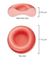
Erythrocytes are small, flexible cells optimized for oxygen and carbon dioxide transport.
Biconcave shape: Increases surface area for gas exchange.
Lack of organelles: Maximizes space for hemoglobin.
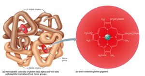
Hemoglobin: Protein that binds oxygen and carbon dioxide reversibly.
Hemoglobin Structure
Hemoglobin is composed of four polypeptide chains (two alpha, two beta), each with a heme group containing iron.
Each iron atom binds one oxygen molecule; thus, each hemoglobin can carry four oxygen molecules.
Hemoglobin also transports some carbon dioxide (as carbaminohemoglobin).
Erythropoiesis: Formation of Red Blood Cells
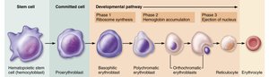
Erythropoiesis is the process of red blood cell production, occurring in red bone marrow.
Begins with hematopoietic stem cells (hemocytoblasts).
Progresses through several stages: proerythroblast, erythroblast, reticulocyte, and mature erythrocyte.
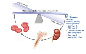
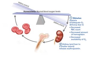
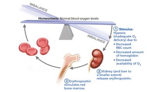
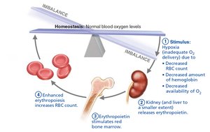
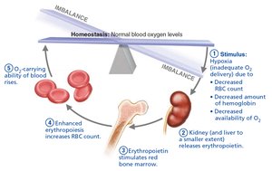
Regulated by erythropoietin (EPO), a hormone released by the kidneys in response to hypoxia.
Regulation of Erythropoiesis
The number of circulating erythrocytes is tightly regulated to balance oxygen delivery and blood viscosity.
Erythropoietin (EPO): Stimulates red blood cell production in response to low oxygen levels.
Dietary requirements: Iron, vitamin B12, and folic acid are essential for erythropoiesis.
Fate and Destruction of Erythrocytes
Red blood cells have a lifespan of 100–120 days. Old cells are removed by macrophages in the spleen, and their components are recycled.
Iron is stored and reused.
Heme is degraded to bilirubin and excreted in bile.
Globin is broken down into amino acids.
Erythrocyte Disorders
Anemia
Anemia is a condition in which the blood's oxygen-carrying capacity is insufficient to support normal metabolism.
Causes: Blood loss, decreased RBC production, or increased RBC destruction.
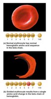
Types: Iron-deficiency anemia, pernicious anemia, renal anemia, aplastic anemia, hemolytic anemias, thalassemia, sickle-cell anemia.
Polycythemia
Polycythemia is an abnormal excess of erythrocytes, increasing blood viscosity and risk of clotting.
Polycythemia vera: Bone marrow cancer.
Secondary polycythemia: Due to increased EPO or low oxygen availability.
Leukocytes (White Blood Cells)
Structure and Function
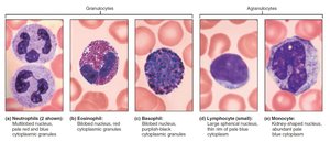
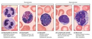
Leukocytes are the only formed elements that are complete cells. They defend the body against infection and disease.
Can leave the bloodstream (diapedesis) and move to sites of infection or injury.
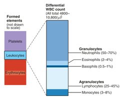
Classified as granulocytes or agranulocytes based on the presence of cytoplasmic granules.
Types of Leukocytes
Neutrophils: Most abundant; phagocytize bacteria.
Eosinophils: Attack parasitic worms; modulate allergic responses.
Basophils: Release histamine; involved in inflammation.
Lymphocytes: T cells (cell-mediated immunity), B cells (antibody production).
Monocytes: Differentiate into macrophages; phagocytize pathogens and debris.
Leukopoiesis: Formation of White Blood Cells
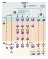
Leukopoiesis is the process of white blood cell formation, regulated by interleukins and colony-stimulating factors.
Originates from hematopoietic stem cells.
Myeloid and lymphoid lineages give rise to different leukocyte types.
Leukocyte Disorders
Leukopenia: Abnormally low WBC count.
Leukemias: Cancers involving overproduction of abnormal WBCs.
Infectious mononucleosis: Viral disease causing excess atypical lymphocytes.
Platelets and Hemostasis
Platelets: Structure and Formation
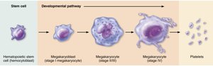
Platelets are cell fragments derived from megakaryocytes and are essential for blood clotting.
Formation (thrombopoiesis) is regulated by thrombopoietin.
Normal concentration: 150,000–400,000 platelets/μl.
Hemostasis: Prevention of Blood Loss
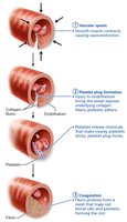
Hemostasis is a rapid, localized process that stops bleeding through three main steps:
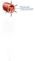
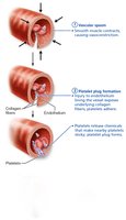
Step 1: Vascular spasm – Vasoconstriction reduces blood flow.
Step 2: Platelet plug formation – Platelets adhere to exposed collagen and aggregate.
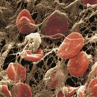
Step 3: Coagulation – Fibrin mesh stabilizes the platelet plug.
Coagulation Pathways
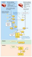
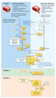
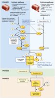
Coagulation involves a cascade of clotting factors leading to the formation of a stable fibrin clot. There are intrinsic and extrinsic pathways, both converging to activate prothrombin to thrombin, which then converts fibrinogen to fibrin.

Clot Retraction and Fibrinolysis
After clot formation, the clot retracts to bring wound edges together, and fibrinolysis dissolves the clot once healing is complete.
Clot retraction: Platelets contract, pulling fibrin threads and squeezing out serum.
Fibrinolysis: Plasminogen is activated to plasmin, which digests fibrin.
Disorders of Hemostasis
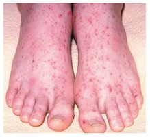
Thromboembolic disorders: Unwanted clot formation (thrombus, embolus).
Bleeding disorders: Impaired clot formation (thrombocytopenia, hemophilia, impaired liver function).
Blood Transfusion and Blood Typing
Blood Groups and Transfusion Compatibility
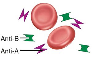
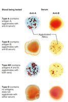
Blood transfusions require careful matching of donor and recipient blood types to prevent immune reactions.
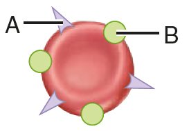
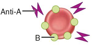
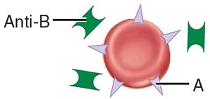
ABO blood groups: Based on presence of A and/or B antigens on RBCs.
Rh blood groups: Based on presence of D antigen (Rh+ or Rh-).
Transfusion reactions can occur if mismatched blood is given, leading to agglutination and hemolysis.
Clinical and Developmental Aspects
Blood Tests
Blood analysis provides valuable diagnostic information, including complete blood count (CBC), differential WBC count, and tests for clotting ability.
Developmental Aspects
Blood cell formation begins in the fetal yolk sac, liver, and spleen, with red bone marrow becoming the primary site by the seventh month. Hemoglobin F in fetuses has a higher affinity for oxygen than adult hemoglobin.
Summary Table: Formed Elements of Blood
Formed Element | Main Function | Relative Abundance | Life Span |
|---|---|---|---|
Erythrocytes | Transport O2 and CO2 | ~99% | 100–120 days |
Leukocytes | Defense against disease | <1% | Hours to years |
Platelets | Clotting | <1% | 5–10 days |
Additional info: This guide covers the essential concepts and clinical relevance of blood as presented in Chapter 17 of a standard Anatomy & Physiology textbook, suitable for exam preparation and foundational understanding.
