 뒤로
뒤로Chapter 17: Blood – Structure, Function, and Clinical Relevance
스터디 가이드 - 스마트 노트
 자료에 맞춘 맞춤형 노트, 핵심 정의, 예시, 맥락을 확장해 제공합니다.
Blood: Characteristics and Functions
Physical Properties of Blood
Blood is a specialized connective tissue with unique physical and functional characteristics essential for homeostasis.
pH: Slightly alkaline, typically 7.35–7.45.
Temperature: About 100.4°F (38°C), slightly higher than body temperature.
Viscosity: Blood is more viscous than water due to cellular and protein content.
Color: Varies from bright red (oxygen-rich) to dark red (oxygen-poor).
Volume: 5–6 L in males; 4–5 L in females.
Functions:
Transport: Oxygen, carbon dioxide, nutrients, wastes, hormones.
Regulation: pH, temperature, fluid balance.
Protection: Immune response, clotting.
Major Components of Whole Blood
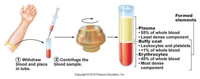
Blood consists of plasma and formed elements, each with distinct roles.
Plasma: The liquid matrix, comprising 55% of blood volume; contains water, proteins (albumin, globulins, fibrinogen), electrolytes, nutrients, and wastes.
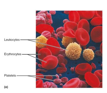
Formed Elements:
Erythrocytes (RBCs): Transport gases.
Leukocytes (WBCs): Immune defense.
Platelets: Clotting.
Hematopoiesis and Blood Cell Formation
Hematopoiesis: The Formation of Blood Cells
Blood cells are produced in the red bone marrow from pluripotent hematopoietic stem cells (hemocytoblasts).
Myeloid Stem Cells: Give rise to RBCs, platelets, monocytes, neutrophils, eosinophils, basophils.
Lymphoid Stem Cells: Give rise to B and T lymphocytes.
Regulatory Factors:
Erythropoietin (EPO): Produced by kidneys; stimulates RBC production.
Thrombopoietin: Produced by liver; stimulates platelet production.
Colony Stimulating Factors & Interleukins: Regulate WBC formation.
Hematocrit
The hematocrit is the percentage of blood volume occupied by RBCs. Normal values: Males ~47%, Females ~42%.
Blood Cell Types
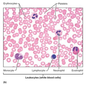
Blood contains several cell types, each with specialized functions.
RBCs: Biconcave, anucleate, filled with hemoglobin.
WBCs: Spherical, nucleated; include granulocytes and agranulocytes.
Platelets: Cytoplasmic fragments essential for clotting.
Erythrocytes (Red Blood Cells)
Structure and Function
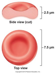
Erythrocytes are specialized for gas transport due to their shape and hemoglobin content.
Shape: Biconcave disc increases surface area for gas exchange.
Anucleate: Mature RBCs lack a nucleus, maximizing space for hemoglobin.
Hemoglobin: Protein responsible for oxygen and carbon dioxide transport.
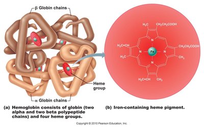
Fate and Destruction of RBCs
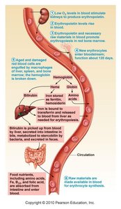
RBCs have a lifespan of about 120 days. Old or damaged RBCs are removed by macrophages, primarily in the spleen and liver.
Iron: Stored as ferritin and hemosiderin; transported by transferrin.
Bilirubin: Waste product from heme breakdown; excreted in bile.
Jaundice: Accumulation of bilirubin causes yellowing of skin and eyes.
Regulation of Erythropoiesis
Erythropoietin Mechanism
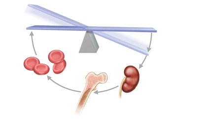
Erythropoiesis is regulated by erythropoietin (EPO), primarily in response to hypoxia (low oxygen levels).
Stimulus: Hypoxia due to decreased RBC count or O2 availability.
Response: Kidneys release EPO, stimulating red bone marrow to increase RBC production.
Homeostasis: Restored when blood O2 levels normalize.
Clinical Aspects of Blood
Complete Blood Count (CBC)
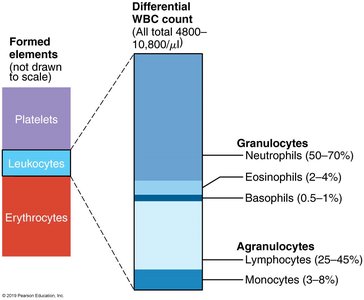
CBC is a common diagnostic test measuring RBCs, WBCs, platelets, hemoglobin, and hematocrit. Differential counts identify specific WBC types.
Anemia
Anemia is a condition of reduced oxygen-carrying capacity of blood. Types include:
Hemorrhagic: Blood loss.
Pernicious: Vitamin B12 deficiency.
Renal: EPO deficiency.
Aplastic: Bone marrow failure.
Hemolytic: RBC destruction (e.g., sickle cell, thalassemia).
Leukocytes (White Blood Cells)
Types and Functions
Leukocytes are divided into granulocytes and agranulocytes, each with distinct roles in immunity.
Granulocytes:
Neutrophils: Phagocytize bacteria; most abundant.
Eosinophils: Kill parasitic worms; involved in allergy/asthma.
Basophils: Release histamine; contain heparin.
Agranulocytes:
Lymphocytes: Mount immune response; B and T cells.
Monocytes: Phagocytosis; develop into macrophages.
Platelets and Hemostasis
Formation of Platelets
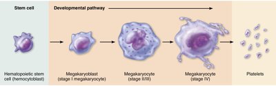
Platelets are formed from megakaryocytes in the bone marrow and are essential for blood clotting.
Hemostasis and Coagulation
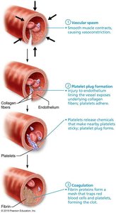
Hemostasis is the process of stopping bleeding, involving three steps:
Vascular Spasm: Vasoconstriction reduces blood flow.
Platelet Plug Formation: Platelets adhere to exposed collagen and release chemicals to attract more platelets.
Coagulation: Blood changes from liquid to gel, forming a fibrin mesh that stabilizes the clot.
Coagulation Pathways
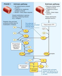
Coagulation involves intrinsic and extrinsic pathways leading to the formation of prothrombin activator, which converts prothrombin to thrombin, and then fibrinogen to fibrin.
Intrinsic Pathway: Initiated by damage inside the vessel.
Extrinsic Pathway: Initiated by external trauma.
Fibrin: Forms the meshwork of the clot.
Fibrinolysis
Fibrinolysis is the process of dissolving the clot after tissue repair, involving plasminogen activation to plasmin, which digests fibrin.
Blood Disorders
Thrombus and Embolus
A thrombus is a stationary blood clot; an embolus is a clot that travels through the bloodstream. Both can cause serious complications.
Bleeding Disorders
Thrombocytopenia: Low platelet count.
Liver Disease: Impaired production of clotting factors.
Hemophilia: Genetic deficiency of clotting factors.
DIC (Disseminated Intravascular Coagulation): Widespread clotting and bleeding.
Restoring Blood Volume
Volume Replacement: Isotonic solutions (normal saline, Ringer’s solution).
RBC Replacement: Packed RBCs or whole blood transfusions.
Shock: Results from >30% blood loss.
Blood Groups and Transfusion
ABO and Rh Blood Groups
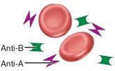
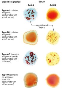
Blood groups are determined by antigens (agglutinogens) on RBC membranes. The ABO and Rh systems are clinically significant.
ABO Groups:
Type A: A antigen, anti-B antibody.
Type B: B antigen, anti-A antibody.
Type AB: A and B antigens, no antibodies (universal recipient).
Type O: No antigens, anti-A and anti-B antibodies (universal donor).
Rh Group: Rh antigen (D); anti-Rh antibodies form only after sensitization.
Hemolytic Disease of the Newborn (Erythroblastosis Fetalis)
Occurs when an Rh-negative mother carries an Rh-positive fetus, leading to antibody formation and fetal RBC destruction in subsequent pregnancies.
Clinical Applications and Review Questions
Blood Transfusion and Testing
Transfusion: Type and crossmatch blood to prevent reactions.
Tests: CBC, blood typing, antibody screening.
Review Questions
James is told he has type A blood. Which ABO antibody/antibodies does he have in his plasma? Which antigens are on his RBCs? Could he donate blood to an AB recipient? Could he receive blood from an AB donor?
Sketch a RBC. Label any antigens present if the blood type is B+. Also write the AB(s) present in the plasma.
A 17 year-old female is brought to the ER with a fever, headache, and stiff neck. You suspect bacterial meningitis. How would you expect her CBC with differential to be abnormal?
Mr. Stars, a 40 year-old teacher from New York, is visiting Colorado to study astronomy. He drives high on a mountain to get a clearer view of the night sky. After a couple of days, he notices he struggles to catch his breath after minimal activity and gets tired quickly. One month later, his symptoms have resolved but he goes to an appointment with his doctor anyway because he is due for an annual physical exam. Would you expect any abnormalities with his CBC? Explain.
Hemoglobin vs. Hematocrit
A young female presents to the ER with severe vaginal bleeding. She is 3 months pregnant. The doctor is concerned with the amount of blood loss. What type of transfusion is most likely to be given to this patient? What blood tests should be ordered before starting the transfusion?
Key Terms and Concepts
Hemoglobin: Oxygen-carrying protein in RBCs.
Hematocrit: Percentage of RBCs in blood.
Plasma: Liquid component of blood.
Platelets: Cell fragments for clotting.
Leukocytes: White blood cells for immunity.
Antigen: Surface marker on RBCs.
Antibody: Protein in plasma targeting foreign antigens.
Important Equations
Hematocrit Calculation:
Oxygen Carrying Capacity:
Summary Table: ABO Blood Groups
Blood Group | RBC Antigens | Plasma Antibodies | Blood That Can Be Received | Universal Status |
|---|---|---|---|---|
A | A | Anti-B | A, O | |
B | B | Anti-A | B, O | |
AB | A, B | None | A, B, AB, O | Universal recipient |
O | None | Anti-A, Anti-B | O | Universal donor |
Summary Table: Formed Elements of Blood
Cell Type | Description | Count (per µL) | Life Span | Function |
|---|---|---|---|---|
Erythrocytes | Biconcave, anucleate | 4–6 million | 100–120 days | Transport O2 and CO2 |
Neutrophils | Multilobed nucleus | 3000–7000 | 6 hours–few days | Phagocytize bacteria |
Eosinophils | Bilobed nucleus, red granules | 100–400 | ~5 days | Kill parasites, allergy/asthma |
Basophils | Bilobed nucleus, purple granules | 20–50 | Few hours–days | Release histamine, heparin |
Lymphocytes | Spherical nucleus | 1500–3000 | Hours–years | Immune response |
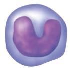
Monocytes | U/kidney-shaped nucleus | 100–700 | Months | Phagocytosis, macrophages |
Platelets | Discoid fragments | 150,000–400,000 | 5–10 days | Clotting |
Additional info: Academic context was added to clarify mechanisms, clinical relevance, and definitions for completeness and exam preparation.
