 뒤로
뒤로Chapter 18: Blood Anatomy and Physiology – Study Guide
스터디 가이드 - 스마트 노트
 자료에 맞춘 맞춤형 노트, 핵심 정의, 예시, 맥락을 확장해 제공합니다.
Blood Anatomy and Physiology
Introduction to Blood
Blood is a specialized connective tissue that plays a vital role in the cardiovascular system, consisting of plasma (the liquid portion) and formed elements (cells and cell fragments). It is essential for transportation, regulation, and protection within the body.
Transportation: Blood carries oxygen, nutrients, hormones, and waste products throughout the body.
Regulation: Maintains pH, body temperature, and fluid balance.
Protection: Provides immunity and prevents blood loss through clotting.
Physical Characteristics of Blood
Blood is more viscous than water, has an alkaline pH (7.35–7.45), and constitutes about 8% of total body weight. Adult males typically have 5–6 L, while females have 4–5 L.
Components of Blood
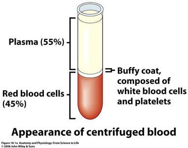
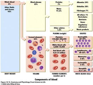
Blood is composed of plasma and formed elements. Plasma makes up 55% of blood, while formed elements (erythrocytes, leukocytes, and platelets) make up 45%.
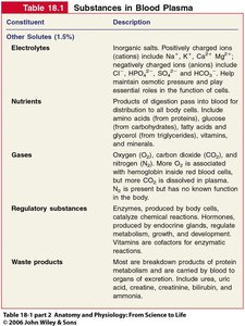
Plasma: Fluid portion containing water, proteins, electrolytes, nutrients, gases, regulatory substances, and waste products.
Formed Elements: Includes erythrocytes (RBCs), leukocytes (WBCs), and platelets.
Blood Plasma
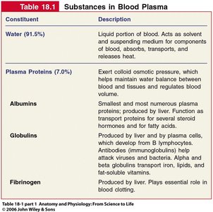
Plasma is primarily water (91.5%), with proteins (7%) and other solutes (1.5%). Plasma proteins include albumin, globulins, and fibrinogen.
Albumin: Maintains osmotic pressure and transports substances.
Globulins: Includes antibodies and transport proteins.
Fibrinogen: Essential for blood clotting.
Formed Elements
Formed elements are living cells and cell fragments, including erythrocytes, leukocytes, and platelets. Hematocrit is the relative percentage of RBCs in total blood volume.
Erythrocytes: Red blood cells responsible for oxygen transport.
Leukocytes: White blood cells involved in immunity.
Platelets: Cell fragments essential for clotting.
Hemopoiesis (Blood Cell Production)
Overview of Hemopoiesis
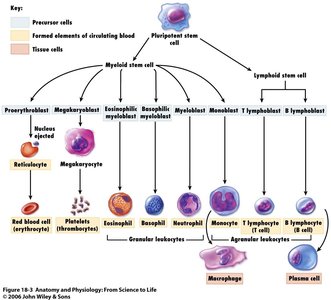
Hemopoiesis is the process by which formed elements develop, primarily occurring in red bone marrow after birth. Pluripotent stem cells differentiate into various blood cells.
Myeloid Line: Forms erythrocytes, most leukocytes, and megakaryocytes (platelet producers).
Lymphoid Line: Forms lymphocytes.
Erythropoiesis
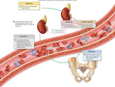
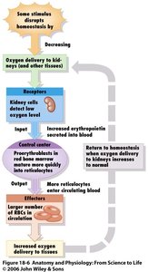
Erythropoiesis is the production of red blood cells, requiring iron, B vitamins, and amino acids. It is regulated by erythropoietin (EPO), a hormone produced mainly in the kidneys.
Stages: Myeloid stem cell → Progenitor cell → Proerythroblast → Erythroblast → Normoblast → Reticulocyte → Erythrocyte
EPO Regulation: Decreased blood oxygen stimulates EPO release, increasing RBC production.
Thrombopoiesis
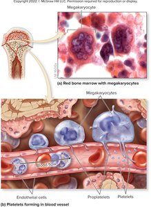
Thrombopoiesis is the production of platelets from megakaryocytes, regulated by thrombopoietin.
Megakaryocytes: Large cells in bone marrow that fragment to form platelets.
Mature Red Blood Cells
Structure and Function
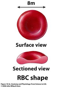
Mature erythrocytes are biconcave, anucleate discs optimized for gas transport. They contain hemoglobin, a protein responsible for carrying oxygen and carbon dioxide.
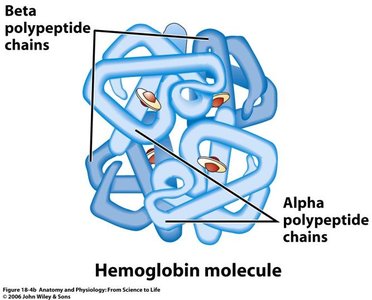
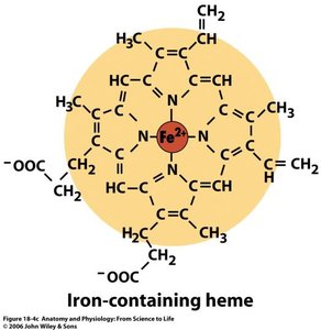
Hemoglobin: Composed of four globin chains (two alpha, two beta), each with a heme group containing iron.
Oxygen Binding: Oxygen binds to iron in heme; each hemoglobin can carry four oxygen molecules.
Carbon Dioxide Transport: CO2 binds to globin protein, not iron.
RBC Life Cycle and Destruction
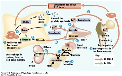
RBCs have a lifespan of about 120 days. Old erythrocytes are phagocytized in the spleen or liver, and their components are recycled.
Globin: Broken down into amino acids for protein synthesis.
Iron: Transported by transferrin, stored as ferritin or hemosiderin, reused for erythropoiesis.
Heme: Converted to biliverdin, then bilirubin, which is excreted in bile, urine, or feces.
Blood Groups
ABO Blood Groups
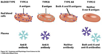
Blood types are determined by the presence of antigens (A, B) on RBCs and corresponding antibodies in plasma. Compatibility is crucial for transfusions.
Type A: A antigen, anti-B antibody
Type B: B antigen, anti-A antibody
Type AB: A and B antigens, no antibodies
Type O: No antigens, both anti-A and anti-B antibodies
Rh Blood Group
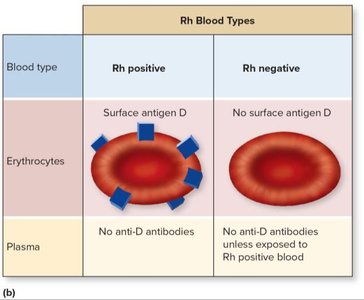
The Rh factor (surface antigen D) determines if blood type is positive or negative. Rh antibodies develop only after exposure to Rh-positive blood in Rh-negative individuals.
Rh Positive: Has antigen D, no anti-D antibodies
Rh Negative: Lacks antigen D, anti-D antibodies only after exposure
Blood Transfusion Compatibility
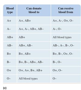
Transfusion reactions occur when incompatible blood is given, leading to agglutination and hemolysis. Proper matching of ABO and Rh types is essential.
Blood Type | Can Donate To | Can Receive From |
|---|---|---|
A+ | A+, AB+ | A+, A-, O+, O- |
A- | A+, A-, AB+, AB- | A-, O- |
AB+ | All blood types | All blood types |
AB- | AB+, AB- | AB-, A-, B-, O- |
B+ | B+, AB+ | B+, B-, O+, O- |
B- | B+, B-, AB+, AB- | B-, O- |
O+ | O+, A+, B+, AB+ | O+, O- |
O- | All blood types | O- |
White Blood Cells (Leukocytes)
Classification and Function
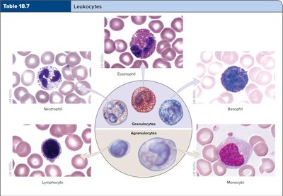
Leukocytes are nucleated cells that defend against pathogens. They are classified as granular (neutrophils, eosinophils, basophils) or agranular (lymphocytes, monocytes).
Granular Leukocytes: Neutrophils, eosinophils, basophils
Agranular Leukocytes: Lymphocytes, monocytes
Granulocytes
Neutrophils: Most numerous, phagocytize pathogens, increase in bacterial infections.
Eosinophils: Combat parasites and allergens, phagocytize antigen-antibody complexes.
Basophils: Release histamine and heparin, involved in allergic responses.
Agranulocytes
Lymphocytes: B cells (antibody production), T cells (cell-mediated immunity), NK cells (attack abnormal cells).
Monocytes: Transform into macrophages, phagocytize microbes and debris.
Platelets
Structure and Function
Platelets are cell fragments derived from megakaryocytes, essential for blood clotting. They circulate for 8–10 days and are stored in the spleen.
Normal Count: 150,000–400,000 per microliter of blood.
Function: Form platelet plugs and release chemicals for clotting.
Hemostasis (Blood Clotting)
Phases of Hemostasis
Hemostasis is the process that stops bleeding, involving three main phases: vascular spasm, platelet plug formation, and coagulation.
Vascular Spasm: Immediate constriction of blood vessels after injury.
Platelet Plug Formation: Platelets adhere to exposed collagen, aggregate, and form a plug.
Coagulation: Formation of a fibrin mesh that traps blood cells and forms a stable clot.
Coagulation Pathways
Coagulation involves intrinsic and extrinsic pathways, both leading to the formation of prothrombinase, which converts prothrombin to thrombin. Thrombin then converts fibrinogen to fibrin, forming the clot.
Intrinsic Pathway: Initiated by damage inside the vessel; involves several clotting factors.
Extrinsic Pathway: Initiated by tissue damage outside the vessel; faster process.
Common Pathway: Both pathways converge, leading to fibrin formation.
Key Equations:
$\text{Prothrombinase} + \text{Ca}^{2+} \rightarrow \text{Thrombin}$
$\text{Thrombin} + \text{Fibrinogen} \rightarrow \text{Fibrin}$
Clot Elimination
Clot retraction and fibrinolysis remove the clot after healing. Actinomyosin contracts the clot, and plasmin degrades fibrin.
Clinical Considerations
Anemia
Anemia is a condition where the percentage of erythrocytes or oxygen-carrying capacity is reduced. Types include aplastic, congenital hemolytic, erythroblastic, hemorrhagic, pernicious, and sickle-cell anemia.
Leukemia
Leukemia is a malignancy in leukocyte-forming cells, leading to abnormal proliferation and decreased erythrocyte and platelet production.
Hemophilia and Clotting Disorders
Hemophilia is a bleeding disorder due to deficiency of clotting factors. Thrombocytopenia is a platelet deficiency, and hypercoagulation is an increased tendency to form clots.
Development and Aging of Blood
Hematopoiesis occurs in most bones in children but is restricted to the axial skeleton in adults. Aging leads to reduced red marrow and increased risk of anemia and leukemia.
Additional info: Academic context and expanded explanations were added to ensure completeness and clarity for exam preparation.
