 뒤로
뒤로Chapter 18: The Cardiovascular System – The Heart (Anatomy & Physiology Study Guide)
스터디 가이드 - 스마트 노트
 자료에 맞춘 맞춤형 노트, 핵심 정의, 예시, 맥락을 확장해 제공합니다.
The Cardiovascular System: The Heart
Overview and Importance
The heart is a central organ in the cardiovascular system, responsible for pumping blood throughout the body via two main circuits: the pulmonary and systemic circuits. Understanding its anatomy and physiology is essential for comprehending how blood is transported, oxygenated, and delivered to tissues.
Heart Structure and Circuits
Pulmonary and Systemic Circuits
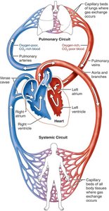
The heart functions as a double pump, with each side serving a distinct circuit:
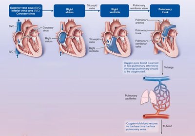
Pulmonary Circuit: Right side of the heart receives oxygen-poor blood and pumps it to the lungs for oxygenation.
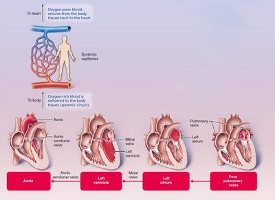
Systemic Circuit: Left side receives oxygen-rich blood from the lungs and pumps it to the rest of the body.
Receiving Chambers: Right atrium (systemic circuit), Left atrium (pulmonary circuit).
Pumping Chambers: Right ventricle (pulmonary circuit), Left ventricle (systemic circuit).
Example: Blood flows from the right atrium to the right ventricle, then to the lungs; from the left atrium to the left ventricle, then to the body.
Size, Location, and Orientation
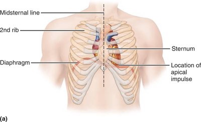
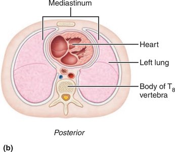
The heart is roughly the size of a fist and located in the mediastinum, between the second rib and fifth intercostal space, on the superior surface of the diaphragm. About two-thirds of the heart lies to the left of the midsternal line, with the apex pointing toward the left hip.
Coverings and Layers of the Heart
Pericardium
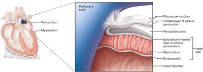
The heart is enclosed in a double-walled sac called the pericardium:
Fibrous Pericardium: Protects, anchors, and prevents overfilling.
Serous Pericardium: Parietal layer lines the fibrous pericardium; visceral layer (epicardium) covers the heart.
Pericardial Cavity: Fluid-filled space between layers reduces friction.
Heart Wall Layers
Epicardium: Visceral layer of serous pericardium.
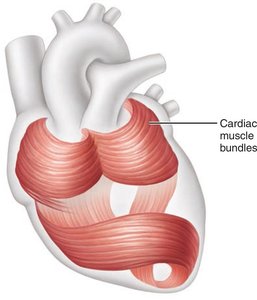
Myocardium: Bulk of the heart wall, composed of contractile cardiac muscle cells arranged in circular and spiral bundles.
Endocardium: Innermost layer, continuous with endothelial lining of blood vessels.
Chambers and Associated Great Vessels
Internal and Surface Features
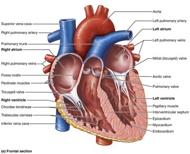
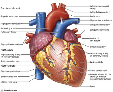
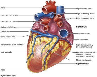
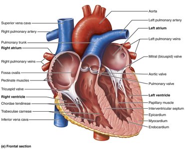
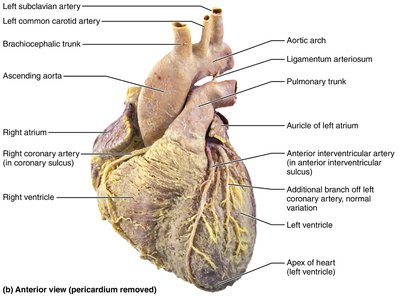
The heart has four chambers: two atria (superior) and two ventricles (inferior). The interatrial septum separates the atria, and the interventricular septum separates the ventricles. Surface features include the coronary sulcus and anterior/posterior interventricular sulci.
Atria: The Receiving Chambers
Auricles: Appendages that increase atrial volume.
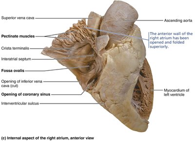
Right Atrium: Receives oxygen-poor blood from the superior/inferior vena cava and coronary sinus.
Left Atrium: Receives oxygen-rich blood from four pulmonary veins.
Pectinate Muscles: Muscle bundles in the atria.
Ventricles: The Discharging Chambers
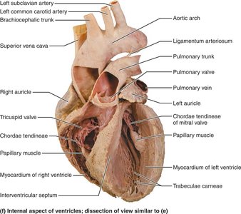
Right Ventricle: Pumps blood into the pulmonary trunk.
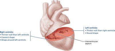
Left Ventricle: Pumps blood into the aorta; has thicker walls due to higher pressure requirements.
Trabeculae Carneae: Irregular ridges of muscle in ventricles.
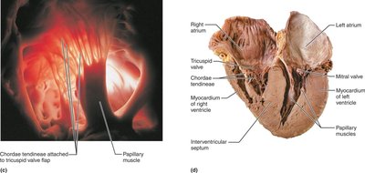
Papillary Muscles: Project into ventricular cavity and anchor valve cusps via chordae tendineae.
Heart Valves
Types and Functions
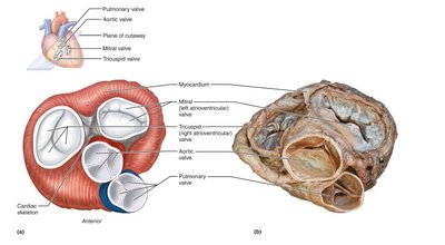
Four heart valves ensure unidirectional blood flow:
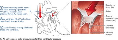
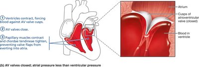
Atrioventricular (AV) Valves: Between atria and ventricles (tricuspid and mitral/bicuspid).
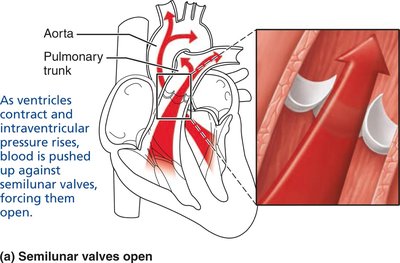
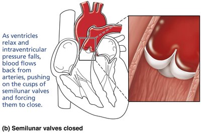
Semilunar (SL) Valves: Between ventricles and major arteries (pulmonary and aortic).
AV Valve Function
Open when atrial pressure exceeds ventricular pressure, allowing blood flow into ventricles.
Close when ventricles contract, preventing backflow into atria.
SL Valve Function
Open when ventricular pressure exceeds arterial pressure, allowing blood to exit the heart.
Close when ventricles relax, preventing backflow from arteries.
Blood Flow Through the Heart
Pathway and Circuit Differences
Blood flows from atria to ventricles, then to either the lungs (pulmonary circuit) or the rest of the body (systemic circuit). Both circuits move equal volumes of blood, but the left ventricle works harder due to higher resistance in the systemic circuit.
Coronary Circulation
Arteries and Veins
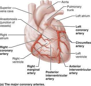
Coronary circulation supplies blood to the heart muscle itself. The left and right coronary arteries branch from the aorta and supply different regions of the heart. Cardiac veins collect deoxygenated blood and return it to the right atrium via the coronary sinus.
Microscopic Anatomy of Cardiac Muscle
Cardiac Myocytes and Intercalated Discs
Cardiac muscle cells are short, branched, and interconnected. Intercalated discs contain desmosomes (for mechanical strength) and gap junctions (for electrical coupling), allowing the myocardium to function as a syncytium.
Comparison: Skeletal vs. Cardiac Muscle
Key Differences
Feature | Skeletal Muscle | Cardiac Muscle |
|---|---|---|
Structure | Striated, long, cylindrical, multinucleate | Striated, short, branched, one or two nuclei |
Gap Junctions | No | Yes |
Contracts as Unit | No | Yes |
T tubules | Abundant | Fewer, wider |
SR | Elaborate | Less elaborate |
Source of Ca2+ | SR only | SR and extracellular fluid |
Pacemaker Cells | No | Yes |
Tetanus Possible | Yes | No |
ATP Supply | Aerobic/anaerobic | Aerobic only |
Intrinsic Conduction System
Pacemaker Cells and Action Potentials
The heart's rhythm is set by pacemaker cells in the sinoatrial (SA) node. The intrinsic conduction system coordinates depolarization and contraction:
SA node → AV node → AV bundle → Bundle branches → Purkinje fibers
Pacemaker potential, depolarization (Ca2+ influx), repolarization (K+ efflux)
Cardiac Cycle
Phases and Heart Sounds
The cardiac cycle includes all events associated with blood flow during one heartbeat:
Systole: Contraction phase
Diastole: Relaxation phase
Heart Sounds: 'Lub' (AV valves close), 'Dup' (SL valves close)
Cardiac Output and Regulation
Stroke Volume and Heart Rate
Cardiac output (CO) is the amount of blood pumped by each ventricle per minute:
Formula:
Stroke Volume (SV):
Ejection Fraction:
Regulation involves preload, contractility, and afterload. Heart rate is influenced by autonomic nervous system, hormones, ions, age, sex, exercise, and temperature.
Clinical Considerations
Homeostatic Imbalances
Pericarditis: Inflammation of pericardium, can lead to cardiac tamponade.
Valve Disorders: Incompetent or stenotic valves cause abnormal heart sounds (murmurs).
Arrhythmias: Irregular heart rhythms, fibrillation, heart block.
Congestive Heart Failure: Inadequate cardiac output, can result from coronary artery disease, hypertension, myocardial infarction, or cardiomyopathy.
Developmental Aspects
Heart Development and Congenital Defects
The heart develops from mesoderm, forming a four-chambered organ by day 35. Fetal structures (foramen ovale, ductus arteriosus) close at birth. Congenital heart defects are common and may involve mixing of blood or narrowed vessels/valves.
Summary Table: Key Differences Between Skeletal and Cardiac Muscle
Feature | Skeletal Muscle | Cardiac Muscle |
|---|---|---|
Structure | Striated, long, cylindrical, multinucleate | Striated, short, branched, one or two nuclei |
Gap Junctions | No | Yes |
Contracts as Unit | No | Yes |
T tubules | Abundant | Fewer, wider |
SR | Elaborate | Less elaborate |
Source of Ca2+ | SR only | SR and extracellular fluid |
Pacemaker Cells | No | Yes |
Tetanus Possible | Yes | No |
ATP Supply | Aerobic/anaerobic | Aerobic only |
Additional info: This study guide covers the anatomy, physiology, and clinical relevance of the heart, suitable for college-level Anatomy & Physiology students. Images included are directly relevant to the anatomical and physiological explanations provided.
