 뒤로
뒤로Chapter 3 – Cells: Structure, Function, and Division
스터디 가이드 - 스마트 노트
 자료에 맞춘 맞춤형 노트, 핵심 정의, 예시, 맥락을 확장해 제공합니다.
Development of the Cell Theory
Overview of Cell Theory
The cell theory is a foundational concept in biology that describes the properties and significance of cells. Cytology, the scientific study of cells, has led to several generalizations:
All organisms are composed of cells and cell products.
The cell is the simplest structural and functional unit of life.
An organism’s structure and functions are due to the activities of its cells.
Cells arise only from preexisting cells.
Common Cell Shapes
Variety of Cell Morphologies
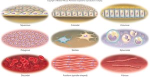
Cells in the human body exhibit a range of shapes, each adapted to specific functions. Understanding these shapes is essential for recognizing cell types in tissues and organs.
Squamous: Thin, flat cells, often found lining surfaces for diffusion and filtration.
Cuboidal: Cube-shaped cells, common in glands and ducts.
Columnar: Tall, column-like cells, specialized for absorption and secretion.
Polygonal: Irregularly angular shapes, often seen in transitional epithelium.
Stellate: Star-shaped, typical of nerve cells.
Spheroidal: Round or oval, such as white blood cells.
Discoid: Disc-shaped, characteristic of red blood cells.
Fusiform: Spindle-shaped, seen in smooth muscle cells.
Fibrous: Long, thread-like, typical of skeletal muscle cells.
The Plasma Membrane
Structure and Components
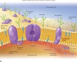
The plasma membrane, also known as the cell membrane, is a selectively permeable barrier that surrounds the cell. Its main components include:
Phospholipids: Form the bilayer, making up 50-75% of the membrane. They provide fluidity and a barrier to most water-soluble substances.
Proteins: Integral and peripheral proteins serve as channels, pumps, receptors, and enzymes.
Glycocalyx: Carbohydrate-rich area on the cell surface, composed of glycoproteins and glycolipids, important for cell recognition, immune response, and tissue compatibility.
Organelles
Internal Structures and Their Functions
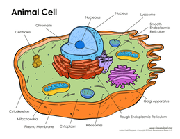
Organelles are specialized structures within cells that perform distinct metabolic tasks:
Cell Membrane: Regulates entry and exit of substances.
Cytoplasm: Intracellular fluid containing organelles.
Cytosol: The fluid portion of the cytoplasm.
Cytoskeleton: Provides structural support and facilitates cell movement and division.
Nucleus: Contains genetic material (DNA).
Ribosomes: Sites of protein synthesis.
Golgi Body: Packages and transports materials.
Mitochondria: Produce ATP through cellular respiration.
Extensions of the Cell Surface
Specialized Structures
Cells may have surface extensions that enhance their functions:
Microvilli: Increase surface area for absorption, especially in the small intestine.
Cilia: Move mucus, trap particles, and serve as sensory receptors; abundant in the respiratory tract.
Flagellum: Whiplike structure for movement; only human sperm have a functional flagellum.
Membrane Transport Mechanisms
Selective Permeability and Transport Types
The plasma membrane is selectively permeable, allowing certain substances to pass while restricting others. Transport mechanisms are classified as passive or active:
Passive mechanisms: Do not require ATP; substances move from high to low concentration (e.g., filtration, simple diffusion, osmosis).
Active mechanisms: Require ATP; substances move from low to high concentration (e.g., active transport, vesicular transport).
Passive Transport
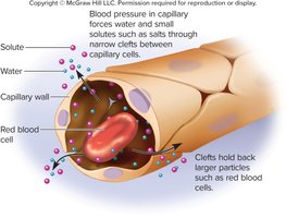
Filtration: Movement of particles through a membrane by physical pressure, important in capillary exchange and kidney function.
Simple Diffusion: Movement of molecules from high to low concentration due to random motion.
Facilitated Diffusion: Diffusion assisted by protein channels; no ATP required.
Osmosis
Osmosis is the net movement of water across a selectively permeable membrane from an area of low solute concentration to high solute concentration. Aquaporins (water channels) facilitate this process. Osmotic imbalances can lead to clinical conditions such as diarrhea, constipation, and edema.
Osmolarity and Tonicity
Osmolarity: The concentration of solutes per liter of solution.
Tonicity: The effect of a solution on cell volume, determined by the concentration of nonpermeating solutes.
Tonicity Types
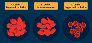
Hypotonic solution: Lower solute concentration outside the cell; water enters, cell swells and may burst (lyse).
Hypertonic solution: Higher solute concentration outside the cell; water leaves, cell shrivels (crenates).
Isotonic solution: Equal solute concentration; no net water movement, cell volume remains stable.
DNA Structure and Function
Genetic Material
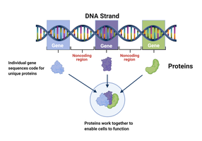
Deoxyribonucleic acid (DNA) is a long, double-helical molecule found in the nucleus. It contains genes, which are segments of DNA coding for specific proteins. Humans have about 20,000 genes distributed across 46 chromosomes.
RNA Structure and Function
Types of RNA and Their Roles
Ribonucleic acids (RNAs) are involved in protein synthesis:
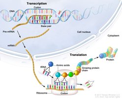
Messenger RNA (mRNA): Carries genetic code from DNA to ribosomes (transcription).
Ribosomal RNA (rRNA): Forms ribosomes, reads mRNA (translation).
Transfer RNA (tRNA): Brings amino acids to ribosomes, assembles proteins according to mRNA code.
The Cell Cycle
Phases of the Cell Cycle
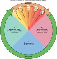
The cell cycle consists of interphase and the mitotic phase:
Interphase: Cell grows, replicates DNA, and prepares for division.
Mitotic phase (Mitosis): Division of the nucleus and cytoplasm to form two new cells.
Functions of the cell cycle include tissue growth, repair, and replacement of dead cells.
Stages of Mitosis
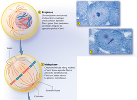
Prophase: Chromosomes condense, nuclear envelope breaks down.
Metaphase: Chromosomes align at the cell's equator.
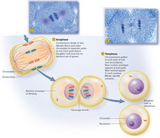
Anaphase: Chromatids separate and move to opposite poles.
Telophase: Nuclear envelopes reform around separated chromosomes.
Cytokinesis: Division of the cytoplasm, resulting in two daughter cells.
DNA Replication and Mutations
DNA Replication
Before cell division, DNA is duplicated during the S phase of interphase to ensure each daughter cell receives a complete set of genetic instructions. This process must be highly accurate to maintain cellular function.
Mutations
Mutations are changes in DNA sequence caused by replication errors or environmental factors (e.g., radiation, chemicals, viruses). Effects range from harmless to causing cell death, cancer, or genetic disorders.
Regulation of the Cell Cycle (Checkpoints)
Cell Cycle Control
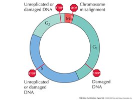
The cell cycle is regulated by checkpoints that ensure proper cell size, DNA integrity, and chromosome alignment before division. Errors that bypass these checkpoints can lead to uncontrolled cell growth or cancer.
Cancer
Causes and Characteristics
Cancer is primarily caused by mutations during DNA replication or exposure to carcinogens (e.g., radiation, chemicals, viruses). It is characterized by uncontrolled cell growth and the potential to invade other tissues (malignancy). Benign tumors are less aggressive and do not metastasize.
Metastasis
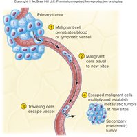
Malignant tumors can spread (metastasize) via the circulatory or lymphatic systems, forming secondary tumors in new locations. Tumors may also stimulate angiogenesis (formation of new blood vessels) to support their growth.
Effects of Cancer
Replacement of functional tissue in vital organs
Invasion of blood vessels, lungs, or brain tissue
Disruption of organ function and nutrient theft
Weakened immunity, increasing susceptibility to infections
Classification of Cancers
Carcinomas: Originate in epithelial tissue
Lymphomas: Originate in lymph nodes
Melanomas: Originate in pigment cells (melanocytes)
Leukemias: Originate in blood-forming tissues
Sarcomas: Originate in bone, connective tissue, or muscle
Oncology is the medical specialty focused on the study and treatment of tumors.
