 뒤로
뒤로Chapter 3: Cells – The Living Units (Anatomy & Physiology Study Notes)
스터디 가이드 - 스마트 노트
 자료에 맞춘 맞춤형 노트, 핵심 정의, 예시, 맥락을 확장해 제공합니다.
Cells: The Smallest Living Units
Cell Theory and Cell Diversity
The cell is the fundamental structural and functional unit of life. The activities of an organism depend on the individual and collective activities of its cells. The structure of each cell is closely related to its function, and all cells arise from preexisting cells, ensuring the continuity of life. Human bodies contain over 250 different types of cells, each varying in size, shape, and subcellular components, which determine their specialized functions.
Cell Theory: All living things are composed of cells; cells are the basic unit of structure and function; new cells arise from existing cells.
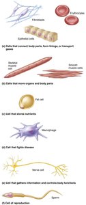
Cell Diversity: Includes cells that connect body parts, move organs, store nutrients, fight disease, gather information, and are involved in reproduction.
Generalized Cell Structure
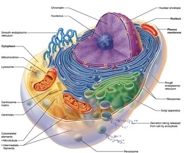
Despite their diversity, all human cells share three basic parts:
Plasma membrane: The flexible outer boundary that separates the cell from its environment.
Cytoplasm: The intracellular fluid containing organelles.
Nucleus: The control center containing DNA.
The Plasma Membrane
Structure and Composition
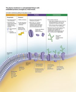
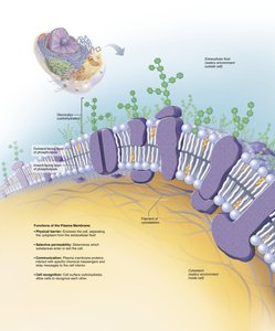
The plasma membrane is a selectively permeable barrier composed of a phospholipid bilayer with embedded proteins, cholesterol, and carbohydrates. This structure is often described as a fluid mosaic model, allowing for flexibility and dynamic interactions.
Phospholipids: Form the basic structure, with hydrophilic heads and hydrophobic tails.
Proteins: Integral and peripheral proteins serve as channels, receptors, enzymes, and anchors.
Cholesterol: Stabilizes membrane fluidity.
Carbohydrates: Contribute to cell recognition and signaling.
Functions of the Plasma Membrane
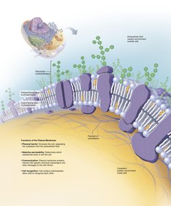
Physical barrier: Separates intracellular and extracellular environments.
Selective permeability: Regulates entry and exit of substances.
Communication: Contains receptors for signal transduction.
Cell recognition: Glycoproteins serve as identification tags.
Membrane Proteins and Their Functions
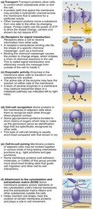
Membrane proteins perform a variety of essential tasks:
Transport: Channels and carriers move substances across the membrane.
Receptors: Bind signaling molecules and initiate cellular responses.
Enzymatic activity: Catalyze reactions at the membrane surface.
Cell-cell recognition: Glycoproteins identify cells to each other.
Intercellular joining: Proteins form junctions between cells.
Attachment: Anchor the membrane to the cytoskeleton and extracellular matrix.
Cell Junctions
Cells are connected by specialized junctions that facilitate communication and maintain tissue integrity:
Tight junctions: Prevent leakage between cells.
Desmosomes: Provide mechanical strength by anchoring cells together.
Gap junctions: Allow direct communication between cells via channels.
Membrane Transport
Passive Membrane Transport
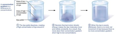
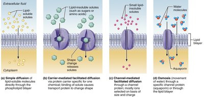
Passive transport does not require energy and relies on the movement of molecules down their concentration gradients. Types include:
Simple diffusion: Movement of small, nonpolar molecules directly through the lipid bilayer.
Facilitated diffusion: Movement of larger or polar molecules via protein channels or carriers.
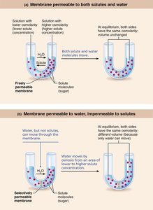
Osmosis: Diffusion of water across a selectively permeable membrane.
Filtration: Movement of water and solutes through a membrane by hydrostatic pressure.
Osmosis and Tonicity
Osmosis is the movement of water across a membrane. Tonicity describes how a solution affects cell volume:
Isotonic: No net water movement; cell size remains constant.
Hypertonic: Water leaves the cell; cell shrinks (crenation).
Hypotonic: Water enters the cell; cell swells and may burst (lysis).
Active Membrane Transport
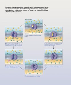
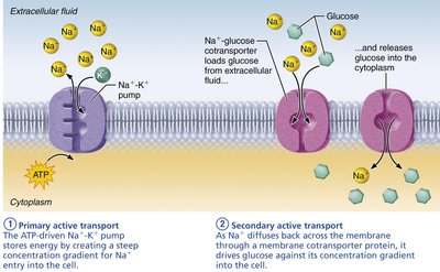
Active transport requires ATP to move substances against their concentration gradients. Two main types:
Primary active transport: Direct use of ATP, e.g., Na+/K+ pump.
Secondary active transport: Indirect use of ATP; uses gradients established by primary active transport to move other substances.
Vesicular Transport
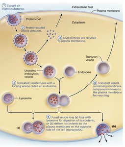
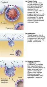
Vesicular transport moves large particles and macromolecules across membranes in vesicles. Types include:
Endocytosis: Uptake of materials into the cell (phagocytosis, pinocytosis, receptor-mediated endocytosis).
Exocytosis: Release of materials from the cell.
Transcytosis: Transport into, across, and out of the cell.
Vesicular trafficking: Movement of substances within the cell.
Resting Membrane Potential
The resting membrane potential is the voltage difference across the plasma membrane in resting cells, primarily established by the movement of potassium (K+) ions. The inside of the cell is negatively charged relative to the outside.
K+ diffusion: Outward movement of K+ creates a negative charge inside the cell.
Na+ diffusion: Inward movement of Na+ slightly offsets the negative charge.
Membrane potential: Typically around -70 mV in neurons.
Cytoplasm and Organelles
Cytoplasm Components
The cytoplasm is the cellular material between the plasma membrane and the nucleus. It consists of:
Cytosol: Gel-like solution containing water, proteins, salts, and sugars.
Inclusions: Insoluble molecules such as glycogen granules, pigments, and lipid droplets.
Organelles: Specialized structures with specific functions, either membranous or nonmembranous.
Mitochondria
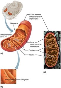
Mitochondria are the cell's power plants, generating ATP through aerobic respiration. They have a double membrane, with the inner membrane folded into cristae. Mitochondria contain their own DNA and can divide independently by fission.
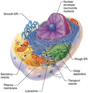
Endoplasmic Reticulum (ER)
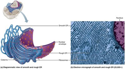
Rough ER: Studded with ribosomes; synthesizes proteins for secretion.
Smooth ER: Lacks ribosomes; involved in lipid metabolism, detoxification, and calcium storage.
Golgi Apparatus
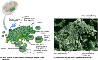
The Golgi apparatus modifies, concentrates, and packages proteins and lipids from the ER. It consists of stacked, flattened membranous sacs (cisternae) and directs the final products to their destinations.
Lysosomes
Lysosomes are spherical organelles containing digestive enzymes. They break down ingested bacteria, viruses, toxins, and worn-out organelles, and play a role in cellular metabolism and autolysis.
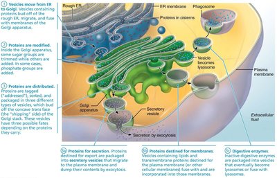
The Endomembrane System
This system includes the ER, Golgi apparatus, secretory vesicles, lysosomes, and the nuclear and plasma membranes. It functions to produce, degrade, store, and export biological molecules, and to degrade harmful substances.
Cytoskeleton
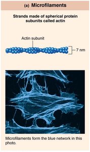
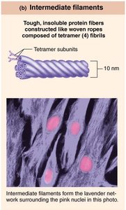
The cytoskeleton provides structural support, facilitates cell movement, and organizes organelles. It consists of:
Microfilaments: Thin filaments of actin involved in cell movement and shape changes.
Intermediate filaments: Tough, rope-like fibers providing mechanical strength.
Microtubules: Hollow tubes of tubulin that determine cell shape and serve as tracks for organelle movement.
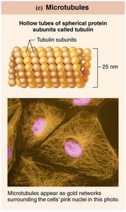
Centrosome and Cellular Extensions
The centrosome is the microtubule organizing center, containing centrioles that form the basis of cilia and flagella. Cilia move substances across cell surfaces, while flagella propel entire cells (e.g., sperm).
The Nucleus and Genetic Control
Nuclear Structure
The nucleus is surrounded by a double-membrane nuclear envelope, which contains nuclear pores for molecular transport. The nucleoplasm fills the nucleus, and the nucleolus is the site of ribosome assembly. The nuclear lamina maintains nuclear shape.
Chromatin and Chromosomes
Chromatin consists of DNA and proteins. During cell division, chromatin condenses to form chromosomes, which protect genetic material during mitosis.
The Cell Cycle and Mitosis
The cell cycle includes interphase (cell growth and DNA replication) and mitotic phase (division of the nucleus and cytoplasm). Mitosis ensures genetic continuity by distributing identical chromosomes to daughter cells.
Protein Synthesis
Protein synthesis involves two main steps:
Transcription: DNA information is copied into messenger RNA (mRNA) in the nucleus.
Translation: mRNA is decoded by ribosomes in the cytoplasm to assemble polypeptides.
There are three types of RNA involved: messenger RNA (mRNA), ribosomal RNA (rRNA), and transfer RNA (tRNA). RNA differs from DNA by having uracil instead of thymine and ribose instead of deoxyribose.
Summary Table: Types of Membrane Transport
Type | Energy Required? | Direction | Examples |
|---|---|---|---|
Simple Diffusion | No | Down gradient | O2, CO2 |
Facilitated Diffusion | No | Down gradient | Glucose, ions |
Osmosis | No | Down water gradient | Water |
Active Transport | Yes (ATP) | Against gradient | Na+/K+ pump |
Vesicular Transport | Yes (ATP) | Varies | Endocytosis, exocytosis |
