 뒤로
뒤로Chapter 3: Cells – The Living Units (Anatomy & Physiology Study Notes)
스터디 가이드 - 스마트 노트
 자료에 맞춘 맞춤형 노트, 핵심 정의, 예시, 맥락을 확장해 제공합니다.
Cells: The Smallest Living Units
Cell Theory
The cell is the fundamental structural and functional unit of all living organisms. The activities of an organism depend on the individual and collective activities of its cells. The structure of each cell is closely related to its function, and all cells arise from preexisting cells, ensuring the continuity of life.
Structural and Functional Unit: All living things are composed of cells, which perform all vital functions.
Complementarity of Structure and Function: The shape and internal structures of a cell determine its biochemical activities.
Cellular Continuity: Cells can only arise from other cells through division.
Cell Diversity
Humans possess over 250 different types of cells, each specialized in size, shape, and internal composition to perform unique functions.
Examples: Muscle cells (movement), nerve cells (information transmission), red blood cells (oxygen transport), fat cells (nutrient storage), macrophages (defense), sperm (reproduction).
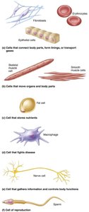
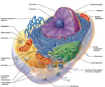
Generalized Cell Structure
Basic Parts of a Human Cell
Despite their diversity, all human cells share three main components:
Plasma Membrane: The flexible outer boundary that separates the cell from its environment.
Cytoplasm: The intracellular fluid containing organelles and inclusions.
Nucleus: The control center containing DNA.
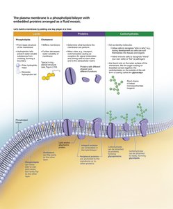
The Plasma Membrane
Structure and Composition
The plasma membrane is a selectively permeable barrier composed of a phospholipid bilayer with embedded proteins, cholesterol, and carbohydrates. It maintains the internal environment of the cell and mediates communication with the external environment.
Phospholipids: Form the basic structure, with hydrophilic heads and hydrophobic tails.
Proteins: Integral and peripheral proteins serve as channels, receptors, enzymes, and anchors.
Cholesterol: Stabilizes membrane fluidity.
Carbohydrates: Contribute to cell recognition and signaling.
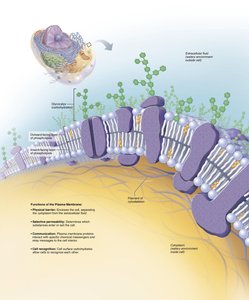
Functions of the Plasma Membrane
Physical Barrier: Separates intracellular and extracellular fluids.
Selective Permeability: Regulates entry and exit of substances.
Communication: Contains receptors for signal transduction.
Cell Recognition: Glycoproteins serve as identification tags.
Membrane Proteins
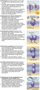
Membrane proteins perform a variety of essential tasks, including transport, signal transduction, enzymatic activity, cell-cell recognition, intercellular joining, and attachment to the cytoskeleton and extracellular matrix.
Cell Junctions
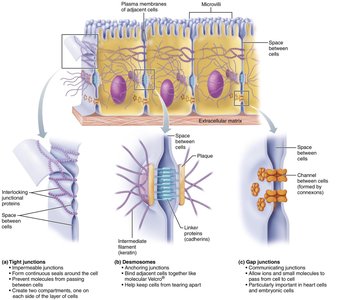
Specialized structures called cell junctions connect adjacent cells, providing communication and structural integrity.
Tight Junctions: Impermeable barriers that prevent leakage between cells.
Desmosomes: Anchoring junctions that provide mechanical stability.
Gap Junctions: Communicating junctions that allow ions and small molecules to pass between cells.
Membrane Transport
Passive Membrane Transport
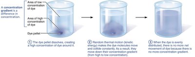
Passive transport does not require cellular energy (ATP). Substances move down their concentration gradients by diffusion, facilitated diffusion, or osmosis.
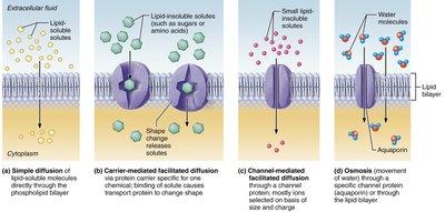
Simple Diffusion: Movement of lipid-soluble molecules directly through the phospholipid bilayer.
Facilitated Diffusion: Movement of molecules via protein carriers or channels (e.g., glucose, ions).
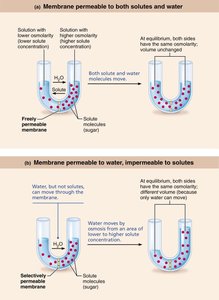
Osmosis: Diffusion of water through a selectively permeable membrane.
Filtration: Movement of water and solutes through a membrane by hydrostatic pressure (mainly in capillaries).
Membrane Permeability and Tonicity
The permeability of the membrane and the tonicity of solutions affect cell volume and function.
Isotonic Solution: Same solute concentration as the cell; no net water movement.
Hypertonic Solution: Higher solute concentration outside the cell; water leaves the cell, causing shrinkage (crenation).
Hypotonic Solution: Lower solute concentration outside the cell; water enters the cell, causing swelling and possible lysis.
Active Membrane Transport
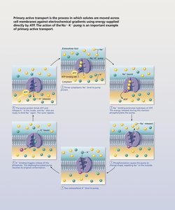
Active transport requires ATP to move substances against their concentration gradients. Two main types are primary and secondary active transport.
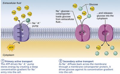
Primary Active Transport: Direct use of ATP to transport molecules (e.g., Na+/K+ pump).
Secondary Active Transport: Indirect use of ATP; uses the energy stored in ion gradients created by primary active transport to drive the movement of other substances.
Vesicular Transport
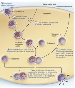
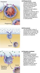
Vesicular transport moves large particles, macromolecules, and fluids across membranes in vesicles. This process requires energy (usually ATP).
Endocytosis: Transport into the cell (includes phagocytosis, pinocytosis, and receptor-mediated endocytosis).
Exocytosis: Transport out of the cell.
Transcytosis: Transport into, across, and out of the cell.
Vesicular Trafficking: Transport from one area or organelle to another within the cell.
Resting Membrane Potential
The resting membrane potential is the voltage difference across the plasma membrane when the cell is at rest, primarily established by the movement of potassium (K+) ions.
Key Role of K+: K+ diffuses out of the cell, creating a negative charge inside. The balance between K+ leaving and being attracted back by the negative charge establishes the resting potential.
Cytoplasm and Organelles
Cytoplasm
The cytoplasm is the cellular material between the plasma membrane and the nucleus. It consists of cytosol (fluid), inclusions (stored nutrients or pigments), and organelles (specialized structures).
Cytosol: Gel-like solution containing water, proteins, salts, and sugars.
Inclusions: Insoluble molecules such as glycogen granules, pigments, and lipid droplets.
Organelles: Metabolic machinery of the cell, each with specialized functions.
Major Organelles
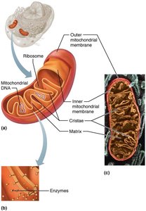
Mitochondria: The "powerhouse" of the cell, producing ATP via aerobic respiration. Contains its own DNA and ribosomes.
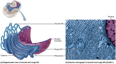
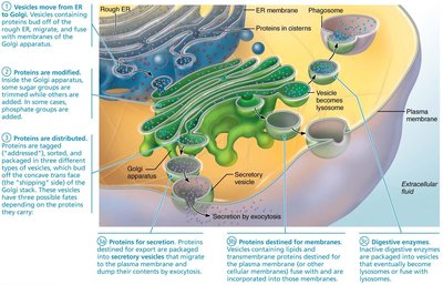
Endoplasmic Reticulum (ER): Network of membranes; rough ER synthesizes proteins, smooth ER synthesizes lipids and detoxifies chemicals.
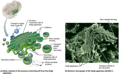
Golgi Apparatus: Modifies, sorts, and packages proteins and lipids for secretion or delivery to other organelles.
Lysosomes: Spherical organelles containing digestive enzymes; break down waste, pathogens, and cellular debris.
Peroxisomes: Contain enzymes for detoxification and lipid metabolism.
Ribosomes: Sites of protein synthesis; can be free or attached to rough ER.
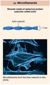
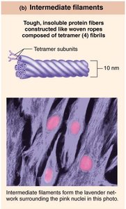
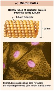
Cytoskeleton: Network of protein filaments (microfilaments, intermediate filaments, microtubules) providing structural support and facilitating movement.
Centrosome and Cellular Extensions
The centrosome is the microtubule organizing center, containing centrioles that play a role in cell division and the formation of cilia and flagella.
Cilia: Short, motile extensions that move substances across cell surfaces.
Flagella: Longer extensions that propel cells (e.g., sperm).
The Nucleus
Structure and Function
The nucleus is the cell's control center, containing genetic material (DNA) and directing cellular activities.
Nuclear Envelope: Double membrane with nuclear pores for molecular exchange.
Nucleolus: Site of ribosomal RNA synthesis.
Chromatin: DNA and protein complex; condenses to form chromosomes during cell division.
Protein Synthesis
Role of RNA
RNA acts as the intermediary between DNA and protein synthesis. There are three main types:
Messenger RNA (mRNA): Carries genetic code from DNA to ribosomes.
Ribosomal RNA (rRNA): Forms part of the ribosome structure.
Transfer RNA (tRNA): Brings amino acids to the ribosome during translation.
Protein synthesis occurs in two main steps:
Transcription: DNA information is coded into mRNA in the nucleus.
Translation: mRNA is decoded at the ribosome to assemble a polypeptide chain.
Key Differences Between DNA and RNA:
RNA contains ribose sugar; DNA contains deoxyribose.
RNA uses uracil instead of thymine.
Additional info: The cell cycle, including mitosis and cytokinesis, ensures the accurate replication and distribution of genetic material to daughter cells. Chromosomes are condensed forms of chromatin, which protect DNA during cell division.
