 뒤로
뒤로Chapter 4: Histology – The Study of Tissues
스터디 가이드 - 스마트 노트
 자료에 맞춘 맞춤형 노트, 핵심 정의, 예시, 맥락을 확장해 제공합니다.
Histology: The Study of Tissues
Introduction to Histology
Histology is the study of the normal structures of tissues, which are groups of structurally and functionally related cells and their external environment. All tissues share two basic components: a discrete population of cells related in structure and function, and an extracellular matrix (ECM) that surrounds these cells.
Cells: The living units performing specific functions.
Extracellular Matrix (ECM): The non-cellular component providing structural and biochemical support.
Types of Tissues
Primary Tissue Types
The human body contains four primary tissue types, each defined by the kind and number of cells, the amount and composition of ECM, and their specific functions:
Epithelial tissues (epithelia): Tightly packed sheets of cells with little to no visible ECM. They cover and line all body surfaces and cavities. Specialized epithelia form glands for secretion (e.g., sweat, saliva, hormones).
Connective tissues: Connect all other tissues to one another. ECM is a prominent feature, and cells are scattered throughout. Functions include binding, support, protection, and transportation of substances.
Muscle tissues: Capable of generating force by contracting. They have little ECM between cells.
Nervous tissues: Capable of generating, sending, and receiving messages. Support cells are also present within a unique ECM.
The Extracellular Matrix (ECM)
Structure and Function of the ECM
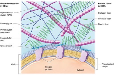
The ECM is a complex network of substances in liquid, gel, or solid form that surrounds the cells of a tissue. It consists of two main components: ground substance and protein fibers.
Ground Substance: The main component of the ECM, containing extracellular fluid (ECF), water, nutrients, ions, and three families of macromolecules:
Glycosaminoglycans (GAGs): Such as chondroitin sulfate and hyaluronic acid; trap water and help resist compression.
Proteoglycans: GAGs bound to a protein core, forming large aggregates that make the ECM firmer and more resistant to compression.
Cell-adhesion molecules (CAMs): Glycoproteins that adhere cells to each other and to the ECM, maintaining tissue structure.
Protein Fibers: Provide tensile strength and structural support. Three main types:
Collagen fibers: Provide resistance to tension and pressure.
Elastic fibers: Allow tissues to stretch and return to their original shape.
Reticular fibers: Form supportive meshworks in organs like the spleen.
Cell Junctions
Types of Cell Junctions
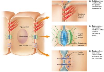
Cell junctions are specialized structures that connect adjacent cells, providing mechanical strength and regulating the passage of substances.
Tight junctions: Make spaces between cells impermeable.
Desmosomes: Increase tissue resistance to mechanical stress.
Gap junctions: Allow small substances to move from one cell to another.
Epithelial Tissues
Functions of Epithelial Tissues
Epithelial tissues cover every internal and external body surface, forming barriers between the body and the external environment. They line organs and cavities and perform several key functions:
Protection: Shield underlying tissues from injury.
Immune defense: Form barriers to prevent microorganism invasion.
Secretion: Form glands that produce substances like hormones and oils.
Transport: Selectively permeable membranes allow substances to cross into other tissues.
Sensation: Rich nerve supply allows detection of environmental changes.
Structural Features of Epithelia
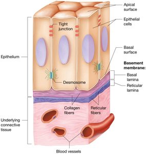
Epithelial tissues are composed of tightly packed cells linked by tight junctions and desmosomes, making them resistant to physical stress. They are avascular (lack blood vessels) and rely on diffusion from underlying tissues for nutrients. The ECM is found beneath the cells in a thin basement membrane, which consists of:
Basal lamina: Collagen fibers and ground substance.
Reticular lamina: Reticular fibers and ground substance, synthesized by underlying connective tissue.
Classification of Epithelia
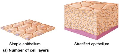
Epithelial tissues are classified by the number of cell layers and the shape of the cells:
Simple epithelia: Single cell layer.
Stratified epithelia: More than one cell layer.
Squamous cells: Flattened shape.
Cuboidal cells: Cube-shaped.
Columnar cells: Tall and elongated.
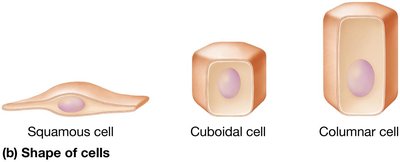
Types of Simple Epithelia
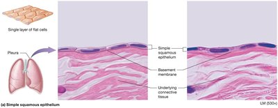
Simple squamous epithelium: Thin, single layer; adapted for rapid diffusion (e.g., air sacs of lungs, kidney tubules, lining blood vessels).
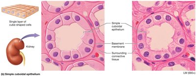
Simple cuboidal epithelium: Single layer of cube-shaped cells; found in kidney tubules and glands.
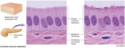
Simple columnar epithelium: Single layer of tall, rectangular cells; often with microvilli or cilia (e.g., small intestine, uterine tubes, respiratory tract).
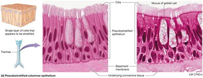
Pseudostratified columnar epithelium: Appears layered due to nuclei at various heights, but is a single layer; found in respiratory tract and nasal cavity.
Transport Across Epithelia
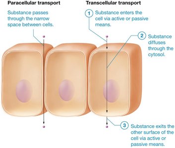
Substances can cross simple epithelia via two routes:
Paracellular transport: Substances leak between cells; limited by tight junctions.
Transcellular transport: Substances pass through the cell, crossing both plasma membranes.
Types of Stratified Epithelia
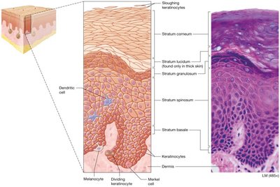
Keratinized stratified squamous epithelium: Apical layers are dead, filled with keratin; found in the outer layers of skin, providing toughness and resistance to friction.
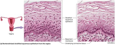
Nonkeratinized stratified squamous epithelium: Apical layers are alive and moist; found in the mouth, throat, esophagus, anus, and vagina.
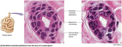
Stratified cuboidal epithelium: Rare; two layers; lines ducts of sweat glands.
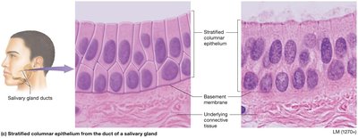
Stratified columnar epithelium: Rare; found in male urethra, cornea, and ducts of certain glands.
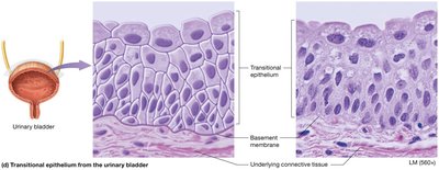
Transitional epithelium: Only in the urinary system; allows tissues to stretch (e.g., bladder, ureters).
Glandular Epithelia
Types and Mechanisms of Secretion
Glands are structures of epithelial origin that synthesize and secrete products. They are classified by shape and how products are released:
Endocrine glands: Secrete hormones directly into the bloodstream; systemic effects.
Exocrine glands: Release products onto epithelial surfaces or into ducts; local effects.
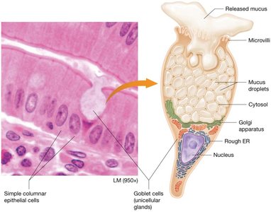
Goblet cells: Most common unicellular exocrine gland; secrete mucus in digestive and respiratory tracts.
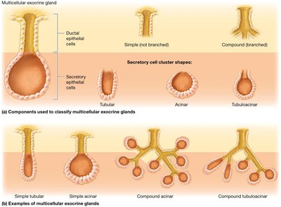
Multicellular exocrine glands: Classified by duct structure (simple or compound) and secretory cell shape (tubular, acinar, tubuloacinar).
Modes of secretion:
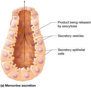
Merocrine: Products released by exocytosis (e.g., salivary and sweat glands).
Holocrine: Cells accumulate product and rupture to release it (e.g., sebaceous glands).
Apocrine: Portions of cytoplasm are pinched off with the product (e.g., mammary glands in some mammals).
Clinical Application: Carcinomas
Epithelial Cancers
Epithelia are more subject to injury and carcinogens (cancer-causing agents) than most other tissues. Carcinoma is the term for epithelial cancer, with common examples including lung adenocarcinoma and basal cell carcinoma of the skin. The basement membrane acts as a barrier to slow the spread of carcinomas, but cancer cells can degrade this barrier to invade other tissues.

Connective Tissue Cells
Immune Cells in Connective Tissue
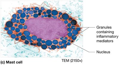
Mast cells: Large resident cells filled with granules of inflammatory mediators (e.g., histamine); release mediators to cause inflammation.
Phagocytes: Immune cells that ingest foreign substances and dead cells; include macrophages and neutrophils.
Additional info: Further details on connective tissue types, muscle tissue, nervous tissue, membranes, and tissue repair are covered in subsequent sections of the chapter.
