 뒤로
뒤로Chapter 4: Histology – The Study of Tissues
스터디 가이드 - 스마트 노트
 자료에 맞춘 맞춤형 노트, 핵심 정의, 예시, 맥락을 확장해 제공합니다.
Histology: The Study of Tissues
Introduction to Tissues
Histology is the study of tissues, which are groups of structurally and functionally related cells and their external environment that together perform common functions. All tissues share two basic components: a discrete population of cells related in structure and function, and the surrounding material called the extracellular matrix (ECM).
Tissue: Group of related cells and their environment performing a common function.
Histology: Study of normal tissue structure.
Extracellular Matrix (ECM): Surrounds and supports cells, providing strength, regulating development, and holding cells in place.
Types of Tissues
Primary Tissue Types
There are four primary tissue types in the human body, each with distinct structures and functions:
Epithelial Tissues: Sheets of tightly packed cells with little ECM; cover and line body surfaces and cavities, and form parts of glands.
Connective Tissues: Connect all other tissues; cells are scattered through ECM; bind, support, protect, and allow transport of substances.
Muscular Tissues: Cells contract to generate force; little ECM.
Nervous Tissues: Neurons generate, send, and receive messages; supported by neuroglial cells with unique ECM.
Extracellular Matrix (ECM)
Components and Functions
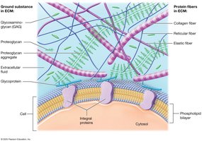
The ECM is composed of substances surrounding the cells in a tissue. It provides strength, directs cell placement, regulates cell activity, and holds cells in position. The ECM consists of two main components: ground substance and protein fibers.
Ground Substance: Gel-like material containing extracellular fluid (ECF), water, ions, nutrients, and macromolecules.
Protein Fibers: Provide tensile strength and support; three types are found in the ECM:
Collagen Fibers: Strong, resistant to tension and pressure.
Elastic Fibers: Made of elastin, allow stretch and recoil.
Reticular Fibers: Thin, form supportive networks.
Cell Junctions
Types of Cell Junctions
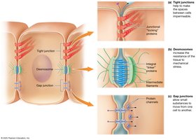
Cell junctions are connections between neighboring cells, formed by integral proteins. They maintain tissue integrity and regulate movement of substances.
Tight (Occluding) Junctions: Seal cells together, preventing passage of macromolecules.
Desmosomes: Distribute mechanical stress, like buttons holding cells together.
Gap Junctions: Protein channels allowing small substances to pass between cells.
Epithelial Tissues
Functions of Epithelial Tissue
Epithelial tissue is found on every external and internal body surface, acting as a barrier between the body and the environment. Its main functions include:
Protection: Shields underlying tissues from injury; produces keratin; rapid mitosis.
Immune Defense: Contains immune cells.
Secretion: Forms glands that produce substances like sweat and hormones.
Transport: Selectively permeable barriers for substance movement.
Sensation: Contains nerves and specialized sensory cells.
Components and Classification of Epithelia
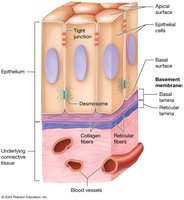
Epithelial tissues are anchored to underlying connective tissue by a basement membrane, which consists of the basal lamina (from epithelial cells) and reticular lamina (from connective tissue). Epithelial cells have an apical surface (facing the lumen or outside), a basal surface (facing the basement membrane), and lateral surfaces (contacting neighboring cells).
Classification by Layers and Cell Shape
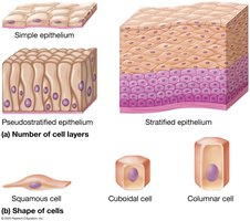
Number of Layers:
Simple Epithelium: Single cell layer.
Stratified Epithelium: Multiple cell layers.
Pseudostratified Epithelium: Appears multilayered but is a single layer.
Cell Shape:
Squamous: Flattened cells.
Cuboidal: Cube-shaped cells.
Columnar: Tall, elongated cells.
Covering and Lining Epithelia
These epithelia are found on inner and outer body surfaces, forming continuous sheets. They are classified based on the number of layers and cell shape.
Simple Epithelia
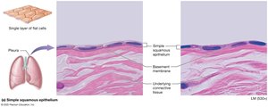
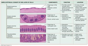
Simple Squamous: Single layer of flat cells; rapid diffusion; found in air sacs of lungs, serous membranes, and blood vessels.
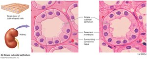
Simple Cuboidal: Single layer of cube-shaped cells; secretion and absorption; found in kidney tubules and glands.
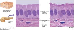
Simple Columnar: Single layer of tall cells; may have microvilli or cilia; found in small intestine, uterine tubes, and glands.
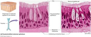
Pseudostratified Columnar: Appears stratified; usually ciliated; contains goblet cells; found in respiratory passages.
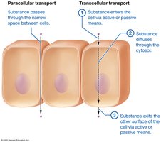
Transport Across Simple Epithelia
Paracellular Transport: Substances pass between cells.
Transcellular Transport: Substances move through cells (enter, diffuse, exit).
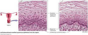
Stratified Epithelia
Stratified Squamous: Multiple layers; protection; nonkeratinized type found in mouth, esophagus, vagina.
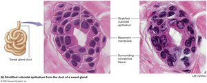
Stratified Cuboidal: Two layers; rare; found in sweat gland ducts.
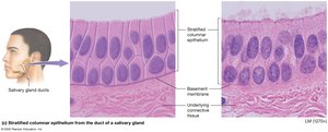
Stratified Columnar: Few layers; rare; found in salivary gland ducts, male urethra, conjunctiva.
Transitional Epithelium: Basal cells cuboidal, apical cells dome-shaped when relaxed, squamous when stretched; found in urinary tract.
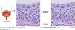
Summary Tables of Epithelial Tissues
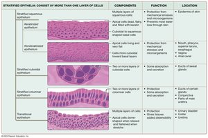
Glandular Epithelia
Types of Glands
Exocrine Glands: Secrete products through ducts to epithelial surfaces; local action.
Endocrine Glands: Secrete hormones directly into the blood; lack ducts; act on distant targets.
Unicellular Exocrine Glands
Goblet Cells: Secrete mucus; found in digestive and respiratory tracts.
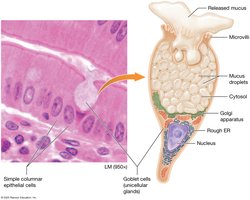
Multicellular Exocrine Glands
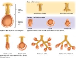
Duct Structure: Simple (unbranched) or compound (branched).
Secretory Shape: Tubular, acinar (spherical), or tubuloacinar (mixed).
Modes of Secretion
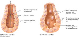
Merocrine: Products released by exocytosis (e.g., sweat glands).
Holocrine: Products released when cell ruptures and dies (e.g., sebaceous glands).
Apocrine: Portions of cytoplasm pinched off with product (e.g., mammary glands).
Connective Tissues
Functions of Connective Tissue
Connecting and Binding: Bind tissue layers and anchor organs.
Support: Bone and cartilage support body weight.
Protection: Bone protects organs; fat and cartilage absorb shock; immune cells present.
Transport: Blood transports substances throughout the body.
Classification of Connective Tissue
Connective Tissue Proper: Loose, dense, reticular, and adipose tissues; widely distributed.
Specialized Connective Tissues: Cartilage, bone, and blood.
Cells of Connective Tissue Proper
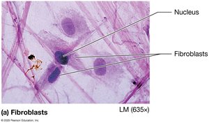
Fibroblasts: Produce fibers and ground substance.
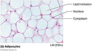
Adipocytes: Store fat.
Mast Cells: Release inflammatory mediators.
Phagocytes: Engulf foreign substances and debris.
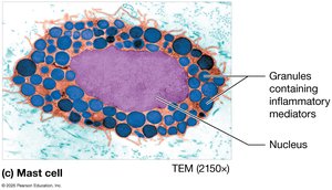
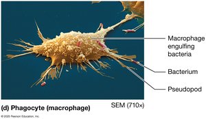
Types of Connective Tissue Proper
Loose (Areolar) Connective Tissue: All three fiber types, supports epithelium, houses blood vessels.
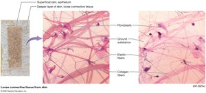
Dense Irregular Connective Tissue: Collagen fibers arranged haphazardly; resists tension in all directions.
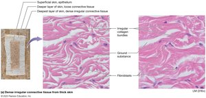
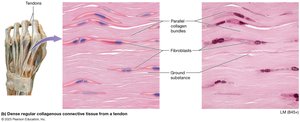
Dense Regular Collagenous Connective Tissue: Parallel collagen bundles; found in tendons and ligaments.
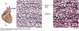
Dense Regular Elastic Connective Tissue: Mostly elastic fibers; found in large blood vessels and some ligaments.
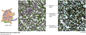
Reticular Tissue: Reticular fibers form supportive networks; found in lymph nodes and spleen.
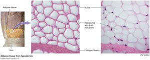
Adipose Tissue: Fat storage, insulation, protection; white and brown types.
Specialized Connective Tissues
Cartilage
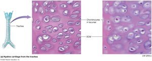
Hyaline Cartilage: Fine collagen fibers; found at bone ends, nose, respiratory tract.
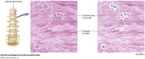
Fibrocartilage: Bundles of collagen; found in intervertebral discs and joints.
Elastic Cartilage: Elastic fibers; found in ear and larynx.
Muscle Tissues
Types of Muscle Tissue
Skeletal Muscle: Voluntary, multinucleate, striated; attached to skeleton for movement.
Cardiac Muscle: Involuntary, branched, single nucleus, striated; found in heart, contains intercalated discs.
Smooth Muscle: Involuntary, non-striated, single nucleus; found in walls of hollow organs and vessels.
Nervous Tissue
Structure and Function
Neurons: Generate and conduct electrical impulses; consist of cell body, axon, and dendrites.
Neuroglial Cells: Support, protect, and nourish neurons; can divide by mitosis.
Membranes
Types of Membranes
Serous Membranes: Line body cavities not open to the exterior; produce serous fluid for lubrication.
Synovial Membranes: Line joint cavities; secrete synovial fluid for joint lubrication.
Mucous Membranes: Line passages open to the exterior; secrete mucus for protection.
Cutaneous Membrane: The skin; protects the body surface.
Tissue Repair
Regeneration and Fibrosis
Regeneration: Damaged cells replaced by the same cell type; restores normal function.
Fibrosis: Damaged cells replaced by dense irregular connective tissue (scar tissue); function may be lost.
The capacity for tissue repair depends on the tissue's ability to undergo mitosis. Epithelial and most connective tissues regenerate well, while cartilage, cardiac muscle, and neurons generally heal by fibrosis.
Summary Table: Epithelial Tissue Types
Type | Structure | Function | Location |
|---|---|---|---|
Simple Squamous | Single layer, flat cells | Diffusion, filtration | Air sacs of lungs, blood vessels |
Simple Cuboidal | Single layer, cube-shaped | Secretion, absorption | Kidney tubules, glands |
Simple Columnar | Single layer, tall cells | Absorption, secretion | Digestive tract, uterine tubes |
Pseudostratified Columnar | Single layer, appears stratified | Protection, secretion | Respiratory tract |
Stratified Squamous | Multiple layers, flat apical cells | Protection | Skin, mouth, esophagus, vagina |
Stratified Cuboidal | Two layers, cube-shaped | Protection, secretion | Sweat gland ducts |
Stratified Columnar | Few layers, tall apical cells | Protection, secretion | Salivary gland ducts, male urethra |
Transitional | Multiple layers, dome-shaped apical cells | Stretch, protection | Urinary bladder, ureters |
