 뒤로
뒤로Chapter 4: Histology – The Study of Tissues
스터디 가이드 - 스마트 노트
 자료에 맞춘 맞춤형 노트, 핵심 정의, 예시, 맥락을 확장해 제공합니다.
Chapter 4: Histology
Introduction to Tissues
Histology is the study of tissues, which are groups of structurally and functionally related cells and their external environment working together to perform common functions. All tissues share two basic components: a discrete population of cells and an extracellular matrix (ECM).
Tissue: Group of related cells and their environment performing a common function.
Histology: Study of normal tissue structure.
Extracellular Matrix (ECM): Non-cellular component surrounding cells, providing structural and biochemical support.
Primary Tissue Types
Epithelial Tissues: Sheets of tightly packed cells with little ECM; cover and line surfaces and form glands.
Connective Tissues: Cells scattered in ECM; bind, support, protect, and transport substances.
Muscular Tissues: Cells contract to generate force.
Nervous Tissues: Cells generate, send, and receive messages.
Module 4.1: The Extracellular Matrix (ECM)
Components and Functions of ECM
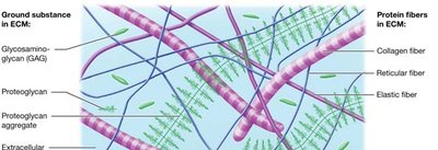
The ECM is composed of substances surrounding the cells in a tissue. It provides strength, directs cell placement, regulates cell development, and holds cells in position. The ECM consists of ground substance and protein fibers.
Ground Substance: Gel-like material containing water, ions, nutrients, and macromolecules.
Protein Fibers: Collagen, elastic, and reticular fibers provide structural support.
Ground Substance Components
Glycosaminoglycans (GAGs): Negatively charged polysaccharides (e.g., chondroitin sulfate, hyaluronic acid).
Proteoglycans: GAGs bonded to a protein core.
Glycoproteins (Cell-Adhesion Molecules, CAMs): Bind cell surface proteins and protein fibers.
Protein Fibers in ECM
Collagen Fibers: Provide resistance to tension and pressure; most abundant protein in the body.
Elastic Fibers: Made of elastin; allow tissues to stretch and recoil.
Reticular Fibers: Thin, short collagen fibers forming supportive scaffolds.
Module 4.1: Cell Junctions
Types of Cell Junctions
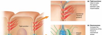
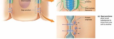
Cell junctions are connections between neighboring cells, formed by integral proteins. They maintain tissue integrity and regulate movement between cells.
Tight Junctions: Integral "locking" proteins create impermeable barriers.
Desmosomes: Integral linker proteins provide mechanical strength by anchoring cells together.
Gap Junctions: Protein channels allow small substances to move between cells.
Module 4.2: Epithelial Tissues
Structure and Function of Epithelial Tissue
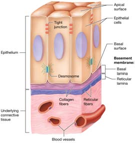
Epithelial tissues are found on every body surface and serve several functions, including protection, immune defense, secretion, transport, and sensation. The ECM of epithelia is located beneath the cells in the basement membrane, which consists of the basal lamina (collagen fibers and ground substance) and the reticular lamina (reticular fibers and ground substance).
Classification of Epithelial Tissue
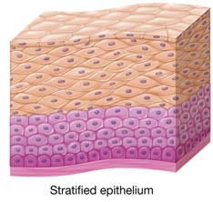
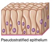
Epithelia are classified by the number of cell layers and the shape of the cells:
Simple Epithelium: One layer of cells.
Stratified Epithelium: Multiple layers of cells.
Cell Shapes: Squamous (flat), cuboidal (cube-shaped), columnar (tall).
Covering and Lining Epithelia
These epithelia are found on inner and outer body surfaces, forming continuous sheets often referred to as membranes.
Simple Epithelial Tissue
Simple epithelia consist of a single layer of cells, allowing for efficient exchange of materials.

Transport Across Simple Epithelium
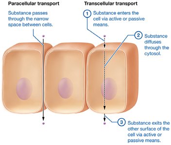
Substances can cross simple epithelia via two main routes:
Paracellular Transport: Through the spaces between cells.
Transcellular Transport: Through the cell, involving entry, diffusion, and exit.
Stratified Epithelial Tissue
Stratified epithelia consist of multiple layers, providing protection against mechanical stress and pathogens.

Homeostatic Imbalances of Epithelial Tissues
Carcinogens: Agents that induce DNA changes and may cause cancer.
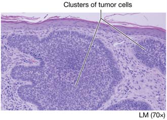
Carcinomas: Cancers of epithelial tissue (e.g., basal cell carcinoma).
Basement Membrane: Can slow the spread of cancer; if not penetrated, cancer is called carcinoma in situ.
Glandular Epithelia
Glands are structures that make and secrete products. They arise from epithelial tissue and are classified as exocrine (secrete via ducts) or endocrine (secrete hormones into blood).
Exocrine Glands: Release secretions to the apical surface via ducts.
Endocrine Glands: Secrete hormones directly into the bloodstream.
Exocrine Gland Structure
Unicellular Glands: Single cells, e.g., goblet cells.
Multicellular Glands: Classified by duct structure and shape of secretory clusters.
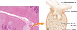
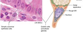
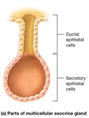
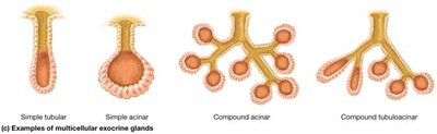
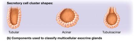
Exocrine Gland Functional Classification
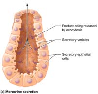
Merocrine Secretion: Products released by exocytosis (e.g., salivary, sweat glands).
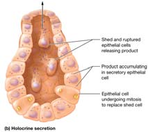
Holocrine Secretion: Products released when cells rupture and die (e.g., sebaceous glands).
Apocrine Secretion: Portions of cytoplasm are pinched off with the product (e.g., mammary glands).
Module 4.3: Connective Tissues
Functions and Classification
Functions: Connecting, binding, support, protection, and transport.
Connective Tissue Proper: Includes loose, dense, reticular, and adipose tissues.
Specialized Connective Tissues: Includes cartilage, bone, and blood.
Cells of Connective Tissue Proper
Fibroblasts: Produce fibers and ground substance.
Adipocytes: Fat cells storing lipids.
Mast Cells: Immune cells with inflammatory mediators.
Phagocytes: Immune cells that engulf foreign substances.

Adipose Tissue
White Adipose: Stores energy, insulates, and cushions organs.
Brown Adipose: Generates heat, especially in infants.
Obesity: Excess adipose tissue; can be hypertrophic (cell enlargement) or hypercellular (increased cell number).
Specialized Connective Tissues: Cartilage
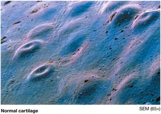
Cartilage is a tough, flexible tissue that absorbs shock and resists tension and compression. It contains chondroblasts (immature cells) and chondrocytes (mature cells in lacunae), and is surrounded by the perichondrium.

Bone or Osseous Tissue
Compact and Spongy Bone: Two types of bone tissue.
Osteoblasts: Build bone matrix.
Osteocytes: Mature bone cells in lacunae.
Osteoclasts: Break down bone matrix.
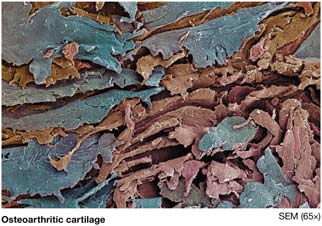
Osteoarthritis
Osteoarthritis is caused by degeneration of hyaline cartilage, leading to joint pain and dysfunction.
Module 4.4: Muscle Tissues
Types of Muscle Tissue
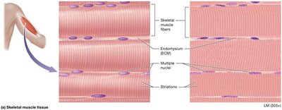
Skeletal Muscle: Voluntary, striated, multinucleated fibers attached to skeleton.
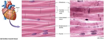
Cardiac Muscle: Involuntary, striated, branched fibers with intercalated discs; found only in the heart.
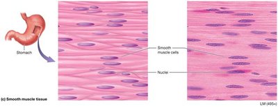
Smooth Muscle: Involuntary, non-striated, spindle-shaped cells; found in walls of hollow organs and vessels.
Module 4.5: Nervous Tissue
Structure and Function
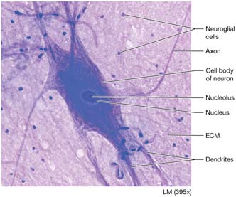
Nervous tissue makes up the brain, spinal cord, and nerves. It consists of neurons (generate and conduct impulses) and neuroglial cells (support and protect neurons).
Module 4.7: Membranes
Types of Membranes
Membranes are thin sheets of tissue lining body surfaces or cavities. They anchor organs, serve as barriers, function in immunity, and secrete substances.
True Membranes: Serous and synovial membranes; do not open to the outside.
Membrane-like Structures: Mucous and cutaneous membranes; open to the outside.
Serous Membranes
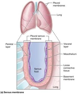
Line pleural, pericardial, and peritoneal cavities; consist of mesothelium, basement membrane, and loose connective tissue. Produce serous fluid to reduce friction.
Synovial Membranes
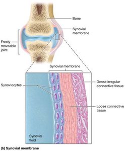
Line freely movable joint cavities; made of synoviocytes and connective tissue. Secrete synovial fluid for joint lubrication.
Mucous Membranes
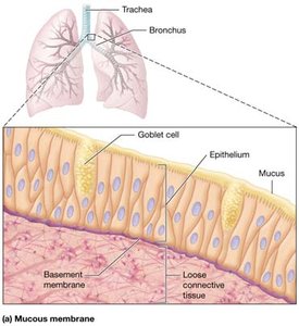
Line body passages opening to the outside; consist of epithelium, basement membrane, lamina propria, and sometimes smooth muscle. Contain goblet cells that secrete mucus.
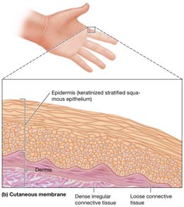
Cutaneous Membrane
The skin; consists of keratinized stratified squamous epithelium and underlying connective tissue.
Module 4.8: Tissue Repair
Regeneration and Fibrosis
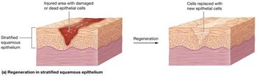
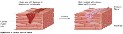
Tissue repair is the process of wound healing, which can occur by regeneration (replacement with original tissue) or fibrosis (replacement with scar tissue).
Regeneration: Replacement of damaged cells with original tissue type.
Fibrosis: Replacement with dense irregular connective tissue (scar tissue).
Capacity for Tissue Repair
Epithelial Tissues: Typically regenerate well due to stem cells.
Connective Tissues: Most regenerate, except cartilage (often heals by fibrosis).
Smooth Muscle: Usually regenerates.
Cardiac and Skeletal Muscle: Generally heal by fibrosis; limited regeneration in skeletal muscle via satellite cells.
Nervous Tissue: Neurons generally do not regenerate.
Other Factors Affecting Tissue Repair
Nutrition: Adequate protein and vitamin C are necessary for collagen synthesis.
Blood Supply: Essential for delivering nutrients and removing waste; poor blood flow impairs healing.
