 뒤로
뒤로Chapter 5: The Integumentary System – Anatomy & Physiology Study Guide
스터디 가이드 - 스마트 노트
 자료에 맞춘 맞춤형 노트, 핵심 정의, 예시, 맥락을 확장해 제공합니다.
The Integumentary System
Overview
The integumentary system is the body's largest organ system, consisting of the skin and its derivatives such as hair, nails, sweat glands, and sebaceous glands. It serves as a protective barrier and plays vital roles in sensation, thermoregulation, and metabolic functions.
Components: Skin, hair, nails, sweat glands, sebaceous (oil) glands
Functions: Protection, regulation of body temperature, sensation, metabolic activities, blood reservoir, excretion
Structure of Skin
Skin Layers
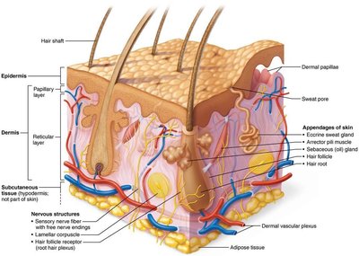
The skin is composed of three main layers, each with distinct functions and structures.
Epidermis: The superficial, avascular layer made of keratinized stratified squamous epithelium.
Dermis: The deeper, vascular layer composed mainly of connective tissue, containing nerves, blood vessels, and glands.
Hypodermis (Subcutaneous layer): Not technically part of the skin, but anchors it to underlying tissues; consists mostly of adipose tissue for shock absorption and insulation.
Epidermis
Cell Types
The epidermis contains four main cell types, each with specialized functions.
Keratinocytes: Produce keratin, the protein that provides protective properties; most abundant cell type.
Melanocytes: Produce melanin pigment, which protects against UV radiation.
Dendritic (Langerhans) cells: Immune cells that patrol the epidermis and activate immune responses.
Tactile (Merkel) cells: Sensory receptors for touch.
Layers of the Epidermis
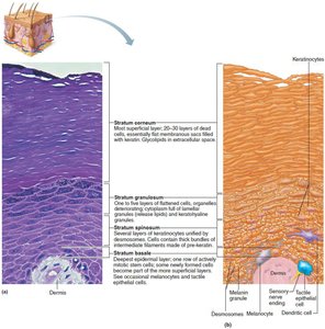
The epidermis is organized into distinct layers (strata), with thick skin containing five layers and thin skin four.
Stratum basale: Deepest layer; single row of stem cells undergoing mitosis. Contains melanocytes.
Stratum spinosum: Several layers thick; contains keratinocytes with prekeratin filaments and dendritic cells.
Stratum granulosum: Four to six layers; cells flatten, keratinization begins, and glycolipids are released for water resistance.
Stratum lucidum: Present only in thick skin; two to three rows of clear, dead keratinocytes.
Stratum corneum: 20–30 layers of dead, keratinized cells; provides protection and prevents water loss.
Dermis
Structure and Layers
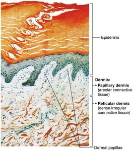
The dermis is a strong, flexible connective tissue layer containing fibroblasts, macrophages, and other cells. It is divided into two layers:
Papillary layer: Superficial areolar connective tissue with dermal papillae, capillary loops, and touch receptors.
Reticular layer: Deep, dense irregular connective tissue; contains collagen and elastic fibers, cutaneous plexus, and pockets of adipose cells.
Dermal Modifications and Skin Markings
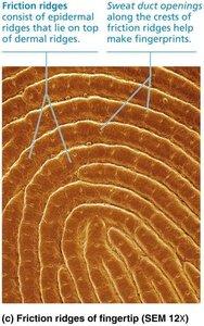
Friction ridges: Found in thick skin; enhance grip and create fingerprints.
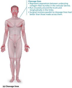
Cleavage (tension) lines: Collagen fibers run parallel to skin surface; important for surgical incisions.
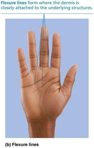
Flexure lines: Dermal folds near joints; visible on hands and feet.
Clinical Aspects
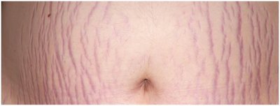
Striae (stretch marks): Dermal tears from extreme stretching.
Blisters: Fluid-filled pockets separating epidermal and dermal layers.
Skin Color
Pigments
Three pigments contribute to skin color:
Melanin: Produced by melanocytes; shields DNA from UV damage.
Carotene: Yellow-orange pigment; accumulates in stratum corneum and hypodermis.
Hemoglobin: Gives pinkish hue to fair skin due to transparency.
Clinical Color Changes
Cyanosis: Blue skin from low oxygenation.
Pallor: Pale skin from anemia or low blood pressure.
Erythema: Redness from fever, inflammation, or allergy.
Jaundice: Yellow skin from liver disorders.
Bruises: Clotted blood beneath skin; color changes as clot breaks down.
Hyperpigmentation: Dark areas may indicate insulin resistance.
Hair
Structure and Function
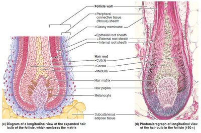
Hair consists of dead, keratinized cells and is produced by hair follicles. Functions include protection, sensation, and regulation of heat loss.
Shaft: Visible part; keratinization complete.
Root: Within scalp; keratinization ongoing.
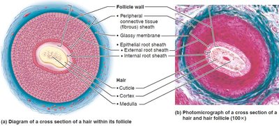
Medulla: Central core.
Cortex: Surrounds medulla.
Cuticle: Outermost layer.
Hair Follicle Structure
Hair bulb: Expanded area at deep end.
Hair follicle receptor: Sensory nerve endings.
Peripheral connective tissue sheath: Derived from dermis.
Glassy membrane: Thickened basal lamina.
Epithelial root sheath: Derived from epidermis.
Hair matrix: Actively dividing area.
Arrector pili: Smooth muscle causing "goose bumps".
Hair papilla: Supplies nutrients.
Types and Growth
Vellus hair: Fine, pale body hair.
Terminal hair: Coarse, long hair (scalp, eyebrows, pubic regions).
Growth: Influenced by nutrition and hormones; follicles cycle between active and regressive phases.
Clinical Aspects
Hirsutism: Excessive hairiness due to androgen excess.
Alopecia: Hair thinning after age 40.
Male pattern baldness: Genetic, influenced by DHT.
Telogen effluvium: Abrupt hair thinning due to stress or illness.
Nails
Structure and Function
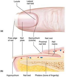
Nails are scale-like modifications of the epidermis containing hard keratin. They protect the distal, dorsal surfaces of fingers and toes.
Free edge: Distal part.
Nail plate: Visible part.
Nail root: Proximal part.
Nail bed: Epidermis under nail plate.
Nail matrix: Responsible for growth.
Nail folds: Skin folds overlapping nail border.
Eponychium: Cuticle.
Hyponychium: Area under free edge.
Lunule: White, thickened area of matrix.
Clinical Aspects
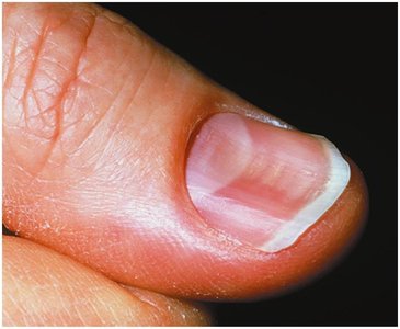
Yellow nails: May indicate respiratory or thyroid disorders.
Thickened yellow nails: Fungal infection.
Koilonychia (spoon nail): Iron deficiency.
Beau’s lines: Severe illness.
Cutaneous Glands
Sweat Glands
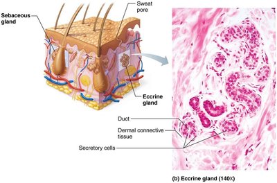
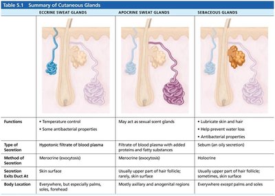
Eccrine (merocrine) glands: Most numerous; thermoregulation; secrete watery sweat.
Apocrine glands: Confined to axillary and anogenital areas; secrete viscous sweat; function as scent glands.
Modified apocrine glands: Ceruminous (earwax) and mammary (milk) glands.
Sebaceous (Oil) Glands
Location: Everywhere except palms and soles.
Function: Secrete sebum (oily, bactericidal); softens skin and hair.
Activity: Stimulated by hormones, especially androgens.
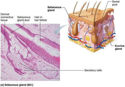
Summary Table: Cutaneous Glands
Gland Type | Function | Secretion | Location |
|---|---|---|---|
Eccrine Sweat Glands | Temperature control, antibacterial | Watery, hypotonic filtrate | Palms, soles, forehead |
Apocrine Sweat Glands | Sexual scent | Viscous, fatty substances | Axillary, anogenital regions |
Sebaceous Glands | Lubricate skin/hair, antibacterial | Oily sebum | Everywhere except palms/soles |
Clinical Aspects
Acne: Infectious inflammation of sebaceous glands.
Whiteheads: Blocked sebaceous glands.
Blackheads: Oxidized secretion.
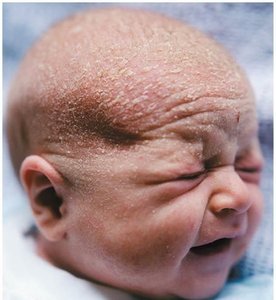
Cradle cap (seborrhea): Overactive sebaceous glands in infants.
Functions of Skin
Protection
The skin acts as a barrier against environmental hazards.
Chemical barrier: Antimicrobial proteins, acid mantle, melanin.
Physical barrier: Keratinized cells, glycolipids, limited permeability.
Biological barrier: Dendritic cells, macrophages, DNA absorbs UV.
Body Temperature Regulation
Insensible perspiration: 500 ml/day under normal conditions.
Sensible perspiration: Up to 12 L/day when overheated.
Cold response: Dermal vessels constrict to conserve heat.
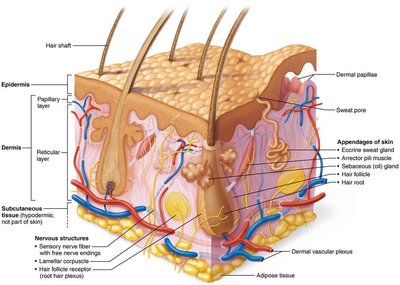
Cutaneous Sensations
Exteroreceptors: Respond to external stimuli (touch, temperature).
Free nerve endings: Sense pain.
Metabolic Functions
Vitamin D synthesis: Needed for calcium absorption.
Keratinocytes: Disarm carcinogens, activate hormones, produce collagenase.
Blood Reservoir
Holds up to 5% of blood volume.
Can be shunted to other organs as needed.
Excretion
Secretes nitrogenous wastes: Ammonia, urea, uric acid.
Sweating: Causes salt and water loss.
Skin Cancer and Burns
Skin Cancer
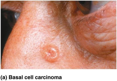
Basal cell carcinoma: Most common, least malignant; arises from stratum basale.
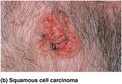
Squamous cell carcinoma: Second most common; arises from keratinocytes of stratum spinosum.
Melanoma: Most dangerous; cancer of melanocytes, highly metastatic.
Burns
First-degree: Epidermal damage only; redness, pain.
Second-degree: Epidermal and upper dermal damage; blisters.
Third-degree: Entire thickness destroyed; gray-white, red, or blackened; no pain (nerve endings destroyed).
Rule of Nines: Used to estimate fluid loss and burn severity.
Critical burns: >25% second-degree, >10% third-degree, or third-degree on face/hands/feet.
Treatment: Debridement, antibiotics, temporary covering, skin grafts.
Developmental Aspects of the Integumentary System
Fetal Development
Lanugo coat: Delicate hairs by 5th–6th month.
Vernix caseosa: Sebaceous gland secretion protecting fetal skin.
Infancy to Adulthood
Skin thickens, accumulates fat.
Gland activity increases, leading to acne.
Optimal appearance in 20s–30s; aging effects after 30.
Aging Skin
Epidermal replacement slows; skin becomes thin, dry, itchy.
Subcutaneous fat and elasticity decrease; wrinkles, cold intolerance.
Increased cancer risk; hair thinning.
Prevention: UV protection, nutrition, hydration, hygiene.
Additional info: Academic context was added to clarify functions, clinical aspects, and developmental changes. Table 5.1 was recreated for clarity and completeness.
