 뒤로
뒤로Compartmentation: Cells & Tissues – Study Notes for Anatomy & Physiology
스터디 가이드 - 스마트 노트
 자료에 맞춘 맞춤형 노트, 핵심 정의, 예시, 맥락을 확장해 제공합니다.
Compartmentation in Human Anatomy
Anatomical Body Compartments
Anatomical compartments are distinct regions within the human body that house specific organs and structures. These compartments are separated by membranes and cavities, which play crucial roles in organ protection and function.
Cranial cavity: Contains the brain.
Thoracic cavity: Houses the heart and lungs, separated by the diaphragm from the abdominopelvic cavity.
Abdominopelvic cavity: Includes abdominal and pelvic organs.
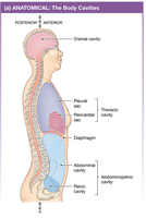
Example: The thoracic cavity contains the pleural sacs (lungs) and pericardial sac (heart).
Lumens: Internal vs. External
A lumen is the interior space of a tubular structure, such as blood vessels or the gastrointestinal tract. Lumens can be lined with endothelium (internal) or epithelium (external).
Internal lumen: Surrounded by endothelium, as in blood vessels.
External lumen: Surrounded by epithelium, as in the digestive tract.
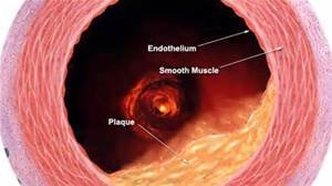
Example: The lumen of the cardiovascular system is considered internal because it is lined by endothelium and not exposed to the external environment.
Functional Body Fluid Compartments
Extracellular Fluid (ECF) vs. Intracellular Fluid (ICF)
The body is divided into two major fluid compartments: extracellular fluid (ECF) and intracellular fluid (ICF). ECF surrounds cells and includes blood plasma and interstitial fluid, while ICF is contained within cells.
ECF: Blood plasma (inside blood vessels) and interstitial fluid (surrounds most cells).
ICF: Fluid inside cells, such as cytoplasm.
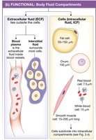
Example: Pericardial fluid is ECF because it is outside cells, in the pericardial sac.
Biological Membranes: Structure & Function
Cell Membrane (Plasma Membrane)
The cell membrane is a selectively permeable barrier that isolates the cell from its environment, regulates exchange, facilitates communication, and provides structural support.
Phospholipid bilayer: Main structural component.
Protein channels & transporters: Regulate movement of substances.
Protein receptors: Enable cell communication.
Anchor for cytoskeleton: Provides structural support.
Fluid Mosaic Model
The fluid mosaic model describes the cell membrane as a dynamic structure composed of a phospholipid bilayer with embedded proteins, cholesterol, and carbohydrates.
Phospholipids: Amphipathic molecules with a polar (hydrophilic) head and non-polar (hydrophobic) tails.
Structures: Bilayer, micelle, and liposome.
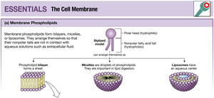
Example: Liposomes can store aqueous digestive enzymes, confining them within the membrane due to its hydrophobic barrier.
Cholesterol in Membranes
Cholesterol is a steroid molecule positioned among the phospholipid fatty acid tails. It is non-polar and hydrophobic, contributing to membrane fluidity and stability.
Membrane Proteins
Membrane proteins are classified as peripheral (attached to the membrane surface) or integral (embedded within the membrane). Integral proteins include transmembrane and lipid-anchored proteins.
Peripheral proteins: Hydrophilic, interact with polar heads.
Integral proteins: Can be hydrophobic or hydrophilic, span the membrane.
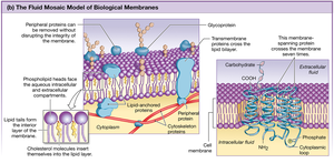
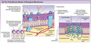
Example: Transmembrane proteins act as channels for ions and molecules.
Concept Map of Cell Membrane Components
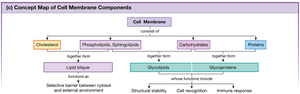
The cell membrane consists of cholesterol, phospholipids, sphingolipids, carbohydrates, and proteins. These components form the lipid bilayer and glycoproteins/glycolipids, which are involved in cell stability, recognition, and immune response.
Intracellular Compartments
Cell Structure Overview
The cell is divided into compartments by the cell membrane. The nucleus contains DNA, and the cytoplasm includes all material between the cell membrane and nucleus.
Cell membrane: Separates inside and outside.
Nucleus: Contains genetic material.
Cytoplasm: Includes organelles and cytosol.
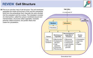
Example: The cytoplasm contains mitochondria, endoplasmic reticulum, and other organelles.
Extracellular & Between Cell Structures
Extracellular Matrix & Cell Junctions
The extracellular matrix is composed of proteins, carbohydrates, and inorganic substances. Cell junctions connect cells and are classified as communicating (gap junctions), occluding (tight junctions), and anchoring (desmosomes).
Gap junctions: Allow direct cell-to-cell communication.
Tight junctions: Block movement of material between cells.
Desmosomes: Anchor cells to each other and to the extracellular matrix.
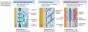
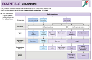
Example: Tight junctions in the intestinal epithelium prevent leakage of digestive contents.
Getting In & Out of the Body: Epithelial Tissue
Epithelial Tissue Anatomy
Epithelial tissue lines all free body surfaces and forms barriers between internal and external environments. The basal lamina (basement membrane) supports epithelial cells. Cell layers and shapes identify epithelial types.
Epithelium: External lining.
Endothelium: Internal lining.
Apical membrane: Faces lumen.
Basolateral membrane: Faces underlying tissue.
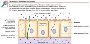
Example: The apical membrane of intestinal epithelial cells faces the gut lumen.
Functions of Epithelial Tissue
Epithelial tissue performs five main functions: exchange, transport, ciliated movement, protection, and secretion. These functions are distributed across various organ systems.
Exchange: Lungs, lining of blood vessels.
Transport: Intestine, kidney.
Ciliated: Nose, trachea, upper airways.
Protection: Skin, lining of mouth.
Secretion: Exocrine glands, stomach, salivary glands.
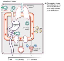
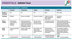
Function | Cell Shape | Special Features | Where Found |
|---|---|---|---|
Exchange | Flattened | Pores between cells permit easy passage | Lungs, lining of blood vessels |
Transporting | Columnar or cuboidal | Tight junctions prevent movement between cells; surface area increased by microvilli | Intestine, kidney |
Ciliated | Columnar or cuboidal | One side covered with cilia to move fluid | Nose, trachea, upper airways, female reproductive tract |
Protective | Flattened in surface layers; polygonal in deeper layers | Cells tightly connected by desmosomes | Skin, lining of mouth, pharynx, esophagus |
Secretory | Columnar or polygonal | Protein-secreting cells filled with secretory granules and extensive RER; steroid-secreting cells contain lipid droplets | Exocrine glands, stomach, salivary glands, sweat glands |
Example: Ciliated epithelium in the respiratory tract moves mucus and trapped particles out of the airways.
