 뒤로
뒤로Endocrine System and Hormone Mapping: Structure, Function, and Regulation
스터디 가이드 - 스마트 노트
 자료에 맞춘 맞춤형 노트, 핵심 정의, 예시, 맥락을 확장해 제공합니다.
Endocrine System Overview
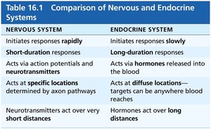
Comparison of Nervous and Endocrine Systems
The nervous and endocrine systems are the primary means of communication and regulation in the human body. While both systems coordinate body functions, they differ in their mechanisms, speed, and duration of action.
Nervous System: Initiates rapid, short-duration responses via action potentials and neurotransmitters, acting at specific locations over short distances.
Endocrine System: Initiates slower, long-duration responses via hormones released into the blood, acting at diffuse locations over long distances.
Nervous System | Endocrine System |
|---|---|
Initiates responses rapidly | Initiates responses slowly |
Short-duration responses | Long-duration responses |
Acts via action potentials and neurotransmitters | Acts via hormones released into the blood |
Acts at specific locations determined by axon pathways | Acts at diffuse locations—targets can be anywhere blood reaches |
Neurotransmitters act over very short distances | Hormones act over long distances |
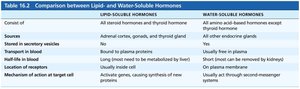
Hormone Classification: Lipid-Soluble vs. Water-Soluble
Hormones are classified based on their solubility, which determines their transport, receptor location, and mechanism of action.
Lipid-Soluble Hormones | Water-Soluble Hormones |
|---|---|
All steroid hormones and thyroid hormone | All amino acid–based hormones except thyroid hormone |
Adrenal cortex, gonads, and thyroid gland | All other endocrine glands |
Bound to plasma proteins | Usually free in plasma |
Long half-life (must be metabolized by liver) | Short half-life (removed by kidneys) |
Inside cell receptors | On plasma membrane receptors |
Activate genes, causing synthesis of new proteins | Usually act through second-messenger systems |
Major Endocrine Organs and Their Histology
Hypothalamus and Pituitary Gland
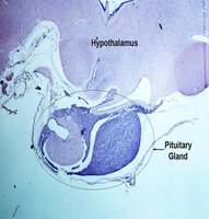
The hypothalamus is a key regulatory center that produces releasing and inhibiting hormones, which control the pituitary gland (hypophysis). The pituitary gland consists of two main lobes: the anterior lobe (adenohypophysis) and the posterior lobe (neurohypophysis).
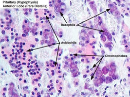
Anterior Lobe: Contains acidophils, basophils, and chromophobes; produces hormones such as GH, TSH, ACTH, FSH, LH, and PRL.
Posterior Lobe: Stores and releases oxytocin and antidiuretic hormone (ADH) produced by the hypothalamus.
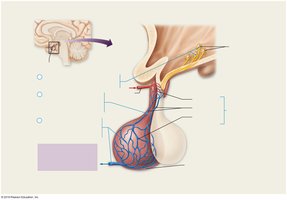
Hypothalamic-Hypophyseal Portal System
This vascular connection allows hypothalamic hormones to reach the anterior pituitary directly, ensuring precise regulation of hormone secretion.
Primary Capillary Plexus in the infundibulum receives hypothalamic hormones.
Hypophyseal Portal Veins transport hormones to the anterior pituitary.
Secondary Capillary Plexus distributes hormones to target cells in the anterior pituitary.
Thyroid Gland
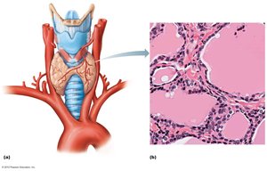
The thyroid gland is located in the anterior neck and consists of two lateral lobes connected by an isthmus. It contains follicles filled with colloid, which stores thyroglobulin, the precursor to thyroid hormones (T3 and T4). Parafollicular cells (C cells) produce calcitonin.
Parathyroid Glands
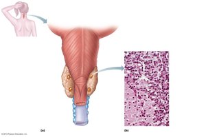
The parathyroid glands are small glands located on the posterior aspect of the thyroid gland. They contain chief (parathyroid) cells that secrete parathyroid hormone (PTH) and oxyphil cells of unknown function.
Adrenal Glands
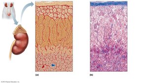
The adrenal glands sit atop the kidneys and consist of an outer cortex and inner medulla. The cortex is divided into three zones: zona glomerulosa (mineralocorticoids), zona fasciculata (glucocorticoids), and zona reticularis (gonadocorticoids). The medulla contains chromaffin cells that secrete epinephrine and norepinephrine.
Pancreas
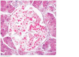
The pancreas has both exocrine and endocrine functions. The endocrine portion consists of pancreatic islets (islets of Langerhans), which contain alpha cells (secrete glucagon) and beta cells (secrete insulin).
Hormones and Their Regulation
Major Hormones and Their Functions
Thyrotropin Releasing Hormone (TRH): Stimulates TSH release from anterior pituitary.
Thyroid Stimulating Hormone (TSH): Stimulates thyroid hormone synthesis and release.
Thyroxine (T4) & Triiodothyronine (T3): Regulate metabolism, growth, and development.
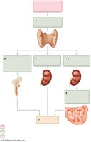
Calcitonin: Lowers blood calcium levels; antagonist to PTH.
Parathyroid Hormone (PTH): Increases blood calcium levels by stimulating osteoclasts, increasing intestinal absorption, and promoting renal reabsorption of calcium.
Gonadotropin Releasing Hormone (GnRH): Stimulates FSH and LH release.
Follicle-Stimulating Hormone (FSH) & Luteinizing Hormone (LH): Regulate gonadal function and gamete production.
Corticotropin Releasing Hormone (CRH): Stimulates ACTH release.
Adrenocorticotropic Hormone (ACTH): Stimulates adrenal cortex to release glucocorticoids.
Growth Hormone Releasing Hormone (GHRH) & Growth Hormone Inhibiting Hormone (GHIH): Regulate GH secretion.
Growth Hormone (GH): Stimulates growth, protein synthesis, and metabolism.
Prolactin (PRL): Stimulates milk production.
Oxytocin: Stimulates uterine contractions and milk ejection.
Antidiuretic Hormone (ADH): Promotes water reabsorption in kidneys.
Aldosterone: Increases sodium reabsorption in kidneys.
Epinephrine & Norepinephrine: Mediate fight-or-flight response.
Insulin: Lowers blood glucose by promoting cellular uptake.
Glucagon: Raises blood glucose by stimulating glycogen breakdown.
Estrogen, Progesterone, Testosterone: Regulate reproductive function.
Hormone Axes and Feedback Loops
Many hormones are regulated through axes involving the hypothalamus, pituitary gland, and target endocrine organs. Negative feedback is the primary mechanism for maintaining homeostasis.
Example: The hypothalamic-pituitary-thyroid axis regulates thyroid hormone levels via TRH, TSH, and thyroid hormones, with negative feedback from T3/T4 to the hypothalamus and pituitary.
Pancreatic Regulation of Serum Glucose
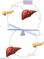
Blood glucose homeostasis is maintained by the antagonistic actions of insulin and glucagon.
Insulin: Released in response to high blood glucose; promotes glucose uptake and storage.
Glucagon: Released in response to low blood glucose; stimulates glycogen breakdown and glucose release from the liver.
Summary Table: Major Endocrine Glands and Hormones
Gland | Hormones Produced | Main Functions |
|---|---|---|
Hypothalamus | TRH, GnRH, CRH, GHRH, GHIH, PIH | Regulates pituitary hormone release |
Anterior Pituitary | GH, TSH, ACTH, FSH, LH, PRL | Growth, metabolism, stress, reproduction, lactation |
Posterior Pituitary | Oxytocin, ADH | Uterine contraction, milk ejection, water balance |
Thyroid | T3, T4, Calcitonin | Metabolism, calcium regulation |
Parathyroid | PTH | Calcium homeostasis |
Adrenal Cortex | Aldosterone, Cortisol, Androgens | Electrolyte balance, stress response, sex hormones |
Adrenal Medulla | Epinephrine, Norepinephrine | Fight-or-flight response |
Pancreas | Insulin, Glucagon | Blood glucose regulation |
Pineal Gland | Melatonin | Regulates circadian rhythms |
Key Terms and Concepts
Target Cells: Cells with specific receptors for a given hormone.
Negative Feedback: A control mechanism in which a change in a physiological variable triggers a response that counteracts the initial change.
Antagonistic Hormones: Hormones that have opposing effects (e.g., insulin and glucagon; calcitonin and PTH).
Additional info:
Some diagrams and tables were inferred to provide a complete overview of endocrine regulation and hormone action.
Histological images were included only when they directly illustrated the structure of the glands discussed.
