 뒤로
뒤로Endocrine System, Blood, and Heart: Mini-Textbook Study Notes
스터디 가이드 - 스마트 노트
 자료에 맞춘 맞춤형 노트, 핵심 정의, 예시, 맥락을 확장해 제공합니다.
Endocrine System
Major Endocrine Glands and Their Hormones
The endocrine system is composed of glands that secrete hormones directly into the bloodstream, regulating various physiological processes. Each gland has specialized cells that produce specific hormones, which act on target organs throughout the body.
Anterior Pituitary (Adenohypophysis): Releases hormones via the hypophyseal portal system.
Corticotropic cells: Secrete Adrenocorticotropic Hormone (ACTH)
Thyrotropic cells: Secrete Thyroid Stimulating Hormone (TSH)
Somatotropic cells: Secrete Growth Hormone (GH)
Gonadotropic cells: Secrete Follicle Stimulating Hormone (FSH) and Luteinizing Hormone (LH)
Prolactin cells: Secrete Prolactin (PRL)
Posterior Pituitary (Neurohypophysis): Hormones are produced in the hypothalamus and transported via the hypothalamic-hypophyseal tract.
Paraventricular nucleus: Produces Oxytocin (OT)
Supraoptic nucleus: Produces Antidiuretic Hormone (ADH)
Pineal Gland: Pinealocytes secrete Melatonin, regulating circadian rhythms.
Thyroid Gland:
Follicular cells: Secrete Triiodothyronine (T3) and Thyroxine (T4)
Parafollicular cells: Secrete Calcitonin
Parathyroid Gland: Parathyroid cells secrete Parathyroid Hormone (PTH), regulating calcium balance.
Pancreas:
Pancreatic Islet β cells: Secrete Insulin
Pancreatic Islet α cells: Secrete Glucagon
Adrenal Cortex:
Zona glomerulosa: Secretes Aldosterone
Zona fasciculata: Secretes Cortisol
Zona reticularis: Secretes Androgens
Adrenal Medulla: Chromaffin cells secrete Epinephrine and Norepinephrine.
Testes: Interstitial cells secrete Testosterone.
Ovaries: Granulosa cells secrete Estrogen and Progesterone.
Other Hormone-Producing Tissues: Adipose (Leptin), GI tract (Gastrin, Secretin), Thymus (Thymosin), Heart (Atrial natriuretic peptide, ANP), Kidneys (Erythropoietin, Renin).
Example: The hypothalamus releases hormones that regulate the anterior pituitary, which in turn controls other endocrine glands, demonstrating the hierarchical nature of endocrine regulation.
Blood
Formed Elements of Blood
Blood consists of plasma and formed elements, which include erythrocytes, leukocytes, and platelets. Each type of cell has a specific function essential for homeostasis.
Erythrocytes (Red Blood Cells): Transport oxygen and carbon dioxide.
Leukocytes (White Blood Cells): Defend the body against infection and foreign substances.
Neutrophils: Phagocytize bacteria.
Eosinophils: Kill parasitic worms; involved in allergy and asthma responses.
Basophils: Release histamine and other mediators of inflammation.
Lymphocytes: Mount immune responses by direct cell attack or via antibodies.
Monocytes: Perform phagocytosis of pathogens and debris.
Platelets (Thrombocytes): Essential for blood clotting.
Example: During an infection, neutrophils rapidly increase in number to combat invading bacteria.
Blood Type Compatibility: Receivers and Donors
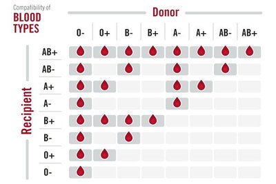
Blood transfusions require compatibility between donor and recipient blood types to prevent immune reactions. The ABO and Rh systems are the primary determinants of compatibility.
Table: Blood Type Compatibility
Key Points:
O- is the universal donor for red blood cells.
AB+ is the universal recipient for red blood cells.
Compatibility depends on the presence or absence of A, B, and Rh antigens.
Example: A patient with blood type A+ can receive blood from A+, A-, O+, and O- donors.
The Heart
Coverings of the Heart
The heart is enclosed in a protective sac called the pericardium, which consists of three layers:
Fibrous pericardium: Outermost, tough connective tissue layer.
Parietal pericardium: Middle serous membrane layer.
Visceral pericardium (Epicardium): Innermost serous layer, directly covering the heart.
Layers of the Heart Wall
Epicardium: Outer layer (same as visceral pericardium).
Myocardium: Thick, muscular middle layer responsible for contraction.
Endocardium: Inner endothelial lining of the heart chambers.
Superficial Landmarks of the Heart
Atrioventricular (Coronary) Sulcus: Separates atria from ventricles.
Anterior and Posterior Interventricular Sulci: Mark the position of the interventricular septum.
Right & Left Auricles: Small, ear-like extensions of the atria.
Apex: Pointed tip of the heart, directed inferiorly and to the left.
Ligamentum Arteriosum: Remnant of fetal ductus arteriosus, connects aorta and pulmonary trunk.
Coronary Circulation
The heart muscle receives its blood supply from the coronary arteries and veins.
Right Coronary Artery: Branches into the marginal artery and posterior interventricular artery.
Left Coronary Artery: Branches into the anterior interventricular artery and circumflex artery.
Cardiac Veins: Great, middle, and small cardiac veins drain into the coronary sinus.
Chambers and Valves of the Heart
Right Atrium: Receives blood from the superior and inferior vena cava; contains pectinate muscles, opening of coronary sinus, and fossa ovalis.
Right Ventricle: Contains tricuspid valve, chordae tendineae, papillary muscles, trabeculae carneae, interventricular septum, pulmonary semilunar valve, pulmonary trunk, and pulmonary artery.
Left Atrium: Receives blood from pulmonary veins.
Left Ventricle: Contains bicuspid (mitral) valve, aortic semilunar valve, and ascending aorta.
Conduction System of the Heart
The heart's intrinsic conduction system coordinates the heartbeat through specialized cardiac muscle cells:
Sinoatrial (SA) Node: Pacemaker of the heart, initiates electrical impulses.
Atrioventricular (AV) Node: Receives impulses from SA node and delays transmission to ventricles.
Atrioventricular (AV) Bundle (Bundle of His): Conducts impulses from AV node to bundle branches.
Right & Left Bundle Branches: Carry impulses through the interventricular septum.
Purkinje Fibers: Distribute impulses throughout ventricular myocardium, causing contraction.
Example: Damage to the SA node can result in arrhythmias, requiring artificial pacemaker intervention.
