뒤로
뒤로Essential Study Notes: Cell Structure, Membrane Transport, Tissues, and Integumentary System
스터디 가이드 - 스마트 노트
 자료에 맞춘 맞춤형 노트, 핵심 정의, 예시, 맥락을 확장해 제공합니다.
Cell Chemistry & Cell Components
Plasma Membrane Structure and Function
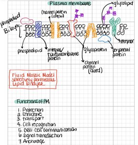
The plasma membrane is a selectively permeable barrier that surrounds the cell, maintaining the internal environment and mediating communication with the external environment. It is described by the Fluid Mosaic Model, which highlights its dynamic and complex structure.
Phospholipid Bilayer: The fundamental structure, composed of two layers of phospholipids with hydrophilic heads facing outward and hydrophobic tails inward.
Integral/Transmembrane Proteins: Span the membrane and are involved in transport and signaling.
Peripheral Proteins: Attached to the membrane surface, often involved in signaling or maintaining cell shape.
Glycoproteins and Glycolipids: Carbohydrate-attached molecules important for cell recognition and signaling.
Channel Proteins: Allow specific molecules or ions to pass through the membrane.
Receptors: Bind signaling molecules and initiate cellular responses.
Functions of the Plasma Membrane:
Protection
Structural support
Transport of substances
Cell recognition
Cell-cell communication
Signal transduction
Anchorage for cytoskeleton and extracellular matrix
Membrane Transport Mechanisms
Cells use various mechanisms to move substances across the plasma membrane, maintaining homeostasis and enabling cellular function.
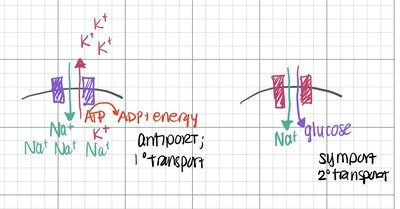
Primary Active Transport (Antiport): Uses ATP to move ions against their concentration gradients in opposite directions (e.g., Na+/K+ pump).
Secondary Active Transport (Symport): Uses the energy from one molecule moving down its gradient to transport another molecule against its gradient in the same direction (e.g., Na+/glucose cotransport).
Key Equations:
For the Na+/K+ pump: per ATP hydrolyzed
Energy & Cell Processes
Cell Cycle and Mitosis
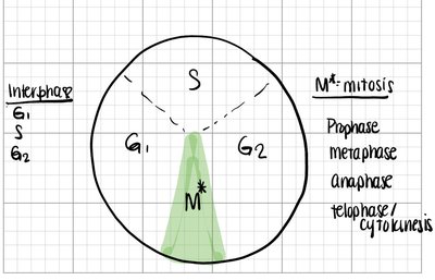
The cell cycle describes the sequence of events in the life of a cell, including growth, DNA replication, and division. Mitosis is the process by which a cell divides to produce two genetically identical daughter cells.
Interphase: Includes G1 (growth), S (DNA synthesis), and G2 (preparation for mitosis).
M Phase (Mitosis): Consists of prophase, metaphase, anaphase, telophase, and cytokinesis.
Mitosis: Chromosome Duplication and Division
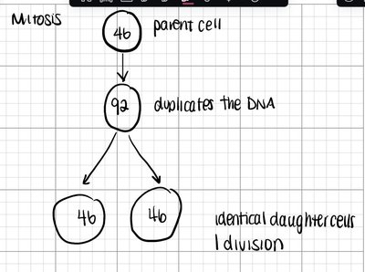
During mitosis, the parent cell duplicates its DNA and divides to form two identical daughter cells, each with the same number of chromosomes as the original cell.
Parent Cell: 46 chromosomes
After DNA Replication: 92 chromatids
Result: Two daughter cells, each with 46 chromosomes
Tissues & Histology
Types of Tissues
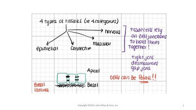
There are four primary tissue types in the human body, each with specialized functions and structures. Tissues rely on cell junctions to maintain integrity and function.
Epithelial Tissue: Covers surfaces and lines cavities.
Connective Tissue: Supports, binds, and protects organs.
Muscular Tissue: Responsible for movement.
Nervous Tissue: Conducts electrical impulses for communication.
Cell Junctions: Tight junctions, desmosomes, and gap junctions hold cells together and allow communication.
Polarity: Epithelial cells can be polarized, with distinct apical (top) and basal (bottom) surfaces.
Epithelial Tissue Classification
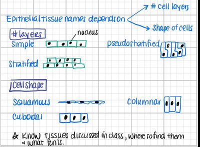
Epithelial tissues are classified based on the number of cell layers and the shape of the cells.
Number of Layers:
Simple: One layer
Stratified: Multiple layers
Pseudostratified: Appears layered but is not
Cell Shape:
Squamous: Flat
Cuboidal: Cube-shaped
Columnar: Tall and column-like
Integumentary System
Overview and Structure
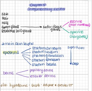
The integumentary system includes the skin, hair, nails, and glands. It serves as the body's first line of defense and plays roles in protection, sensation, and thermoregulation.
Main Skin Layers:
Epidermis: Outermost layer, subdivided into strata (corneum, lucidum, granulosum, spinosum, basale)
Dermis: Deeper layer, includes papillary and reticular regions
Hypodermis (Subcutaneous): Not always considered part of the skin; contains adipose and areolar connective tissue
Glands:
Sudoriferous (sweat) glands: Eccrine (most numerous), apocrine (axillary/genital areas)
Sebaceous (oil) glands
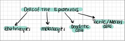
Cells of the Epidermis
The epidermis contains several specialized cell types, each with distinct functions:
Keratinocytes: Produce keratin, a protein that provides strength and waterproofing.
Melanocytes: Produce melanin, the pigment responsible for skin color and UV protection.
Dendritic (Langerhans) Cells: Immune cells that detect and fight pathogens.
Tactile (Merkel) Cells: Sensory receptors for touch.