 뒤로
뒤로Foundations: The Cell – Structure, Function, and Life Cycle
스터디 가이드 - 스마트 노트
 자료에 맞춘 맞춤형 노트, 핵심 정의, 예시, 맥락을 확장해 제공합니다.
Introduction to Cellular Anatomy
Cells are the fundamental structural and functional units of all living organisms. In human anatomy, understanding the structure and function of cells is essential for grasping how tissues, organs, and systems operate. There are two main types of cells in the human body: sex cells (germ cells: sperm and oocytes) and somatic cells (all other body cells).
Cell Structure Overview
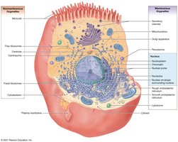
The cell is composed of three primary components: the plasma membrane, cytoplasm (including cytosol and organelles), and various organelles (membranous and nonmembranous). Each component plays a specific role in maintaining cellular function and integrity.
Plasma Membrane
Structure and Function
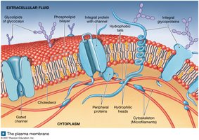
The plasma membrane is a selectively permeable barrier that surrounds the cell, maintaining the internal environment and mediating communication with the external environment. Its four major functions are:
Physical barrier: Separates the cell from its surroundings.
Regulation of exchange: Controls entry and exit of substances.
Sensitivity: Responds to changes in the extracellular environment.
Cell-to-cell communication and adhesion: Facilitates interaction and structural support among cells.
Components of the Plasma Membrane
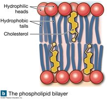
Phospholipid bilayer: Composed of hydrophilic heads facing outward and hydrophobic tails facing inward, forming a double layer that is the primary structure of the membrane.
Proteins:
Peripheral proteins: Attached to the inner or outer surface.
Integral (transmembrane) proteins: Span the membrane; may form channels or act as receptors.
Glycolipids and glycoproteins: Present on the outer layer, forming the glycocalyx, which is involved in cell recognition and signaling.
Sterols (e.g., cholesterol): Stabilize membrane structure.
Membrane Permeability and Transport Mechanisms
Types of Permeability
Impermeable: No substances can cross.
Freely permeable: All substances can cross without restriction.
Selectively permeable: Only certain substances can cross; most biological membranes are selectively permeable.
Passive Transport Processes
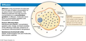
Diffusion: Movement of molecules from high to low concentration (down a concentration gradient). No energy required.
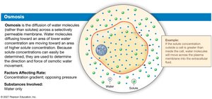
Osmosis: Diffusion of water molecules across a selectively permeable membrane from high to low water concentration.
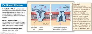
Facilitated diffusion: Passive transport of solutes (e.g., glucose, amino acids) via carrier proteins. Rate depends on concentration gradient, solute size/charge, temperature, and number of carriers.
Active Transport Processes
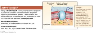
Active transport: Movement of solutes against their concentration gradient using carrier proteins and ATP (e.g., Na+/K+ pump).
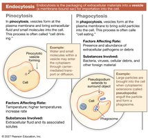
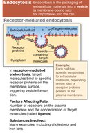
Endocytosis: Uptake of materials into the cell via vesicles.
Pinocytosis: "Cell drinking" – uptake of fluids and small molecules.
Phagocytosis: "Cell eating" – uptake of large particles or pathogens.
Receptor-mediated endocytosis: Selective uptake of specific molecules via receptor binding.
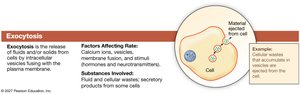
Exocytosis: Release of intracellular materials to the extracellular space via vesicle fusion with the plasma membrane.
Cytoplasm and Its Components
The cytoplasm includes all material inside the cell except the nucleus. It consists of cytosol (intracellular fluid) and organelles (specialized structures performing specific functions).
Cytosol: Contains high K+, low Na+, high protein, small carbohydrates, and inclusions (insoluble substances).
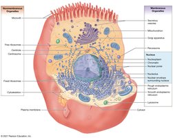
Organelles: Divided into nonmembranous and membranous types.
Nonmembranous Organelles
Cytoskeleton: Provides structural support and facilitates movement. Composed of:
Microfilaments: Actin protein; anchor and move cell structures.
Intermediate filaments: Provide strength and stabilize organelles (e.g., neurofilaments in nerves).
Thick filaments: Myosin protein; abundant in muscle cells for contraction.
Microtubules: Main cytoskeletal component; move organelles, form centrioles, cilia, and flagella.
Centrioles: Organize microtubules during cell division.
Cilia: Move fluids across cell surfaces.
Flagella: Propel sperm cells.
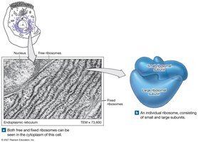
Ribosomes: Sites of protein synthesis; can be free in cytosol or attached to endoplasmic reticulum.
Membranous Organelles
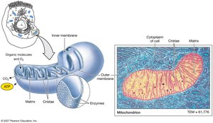
Mitochondria: Double-membraned; produce ATP via aerobic respiration.
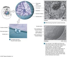
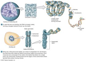
Nucleus: Control center; contains DNA, nucleoli, and chromatin/chromosomes.
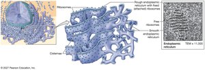
Endoplasmic Reticulum (ER): Network of membranes; two types:
Rough ER (RER): With ribosomes; synthesizes proteins.
Smooth ER (SER): Synthesizes lipids, steroids, carbohydrates; stores Ca2+; detoxifies toxins.
Golgi Apparatus: Stack of cisternae; modifies, sorts, and packages proteins and lipids for secretion or delivery to other organelles.
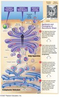
Lysosomes: Contain digestive enzymes; break down waste, pathogens, and cellular debris; involved in autolysis.
Peroxisomes: Contain catalase; neutralize toxins, especially in liver cells, by converting hydrogen peroxide to water and oxygen.
The Cell Life Cycle
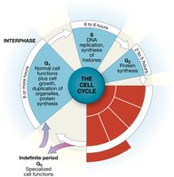
Cell reproduction involves a series of events that ensure genetic material is accurately duplicated and distributed. The main phases are interphase (cell growth and DNA replication) and mitosis (nuclear division), followed by cytokinesis (cytoplasmic division).
Interphase
G1 phase: Cell growth, organelle duplication, and protein synthesis.
S phase: DNA replication and histone synthesis.
G2 phase: Protein synthesis and preparation for mitosis.
G0 phase: Resting phase; cells not preparing to divide.
Mitosis
Mitosis is the process of nuclear division in somatic cells, ensuring each daughter cell receives an identical set of chromosomes. The stages are:
Prophase: Chromatin condenses into visible chromosomes; nuclear envelope dissolves; spindle forms.
Metaphase: Chromosomes align at the cell's equator.
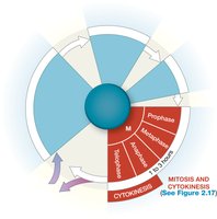
Anaphase: Sister chromatids separate and move to opposite poles.
Telophase: New nuclear membranes form around each set of chromosomes; cell prepares to divide.
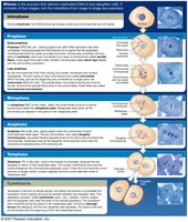
Cytokinesis: Division of the cytoplasm, resulting in two genetically identical daughter cells.
Summary Table: Membranous vs. Nonmembranous Organelles
Nonmembranous Organelles | Membranous Organelles |
|---|---|
Cytoskeleton (microfilaments, intermediate filaments, thick filaments, microtubules) | Mitochondria |
Centrioles | Nucleus |
Cilia | Endoplasmic Reticulum (RER, SER) |
Flagella | Golgi Apparatus |
Ribosomes | Lysosomes |
Peroxisomes |
Additional info: This guide covers the essential structures and functions of the cell, including membrane dynamics, organelle roles, and the cell cycle, as foundational knowledge for further study in anatomy and physiology.
