 뒤로
뒤로Leukocytes, Thrombocytes, and Hemostasis: Blood Defense, Repair, and Typing
스터디 가이드 - 스마트 노트
 자료에 맞춘 맞춤형 노트, 핵심 정의, 예시, 맥락을 확장해 제공합니다.
Leukocytes, Thrombocytes, and Hemostasis
Overview of Blood Defense and Repair
Blood plays a critical role in defense, repair, and maintaining homeostasis. Leukocytes (white blood cells) defend against pathogens, thrombocytes (platelets) mediate clotting, and plasma proteins enable blood typing and compatibility.
Leukocytes: Structure, Classification, and Function
Classification of Leukocytes
Leukocytes are nucleated cells found in blood at concentrations of 4,800–10,800/µL. They are classified based on the presence or absence of cytoplasmic granules:
Granulocytes: Contain visible granules and have lobed nuclei. Types include neutrophils, eosinophils, and basophils.
Agranulocytes: Lack visible granules and have spherical or kidney-shaped nuclei. Types include lymphocytes and monocytes.
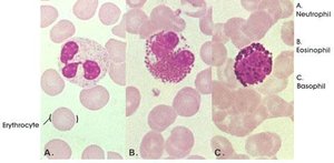
The Granulocyte Trio
Neutrophils (50–70%): Multi-lobed nucleus (3–6 lobes). Active phagocytes that target bacteria and form antimicrobial respiratory bursts.
Eosinophils (2–4%): Bilobed nucleus with coarse red granules. Release enzymes to attack parasites and modulate allergic responses.
Basophils (0.5–1%): U- or S-shaped nucleus hidden by dark granules containing histamine, a potent inflammatory vasodilator.
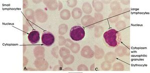
The Agranulocyte Defenders
Lymphocytes (25–45%): Large nucleus occupies most of the cell. Essential for adaptive immunity. T cells attack infected cells; B cells produce antibodies.
Monocytes (3–8%): Largest WBCs with kidney-shaped nuclei. Become tissue macrophages that phagocytose viruses, bacteria, and debris.
Leukopoiesis: Formation of Leukocytes
All blood cells originate from hematopoietic stem cells (hemocytoblasts) in red bone marrow. Leukocyte production is regulated by:
Interleukins (ILs)
Colony-Stimulating Factors (CSFs)
These signals direct precursor cells to differentiate into either the lymphoid line (lymphocytes) or myeloid line (all other formed elements).
Thrombocytes (Platelets): Structure and Function
Platelet Structure and Origin
Platelets are not true cells but cytoplasmic fragments enclosed by a plasma membrane. They are formed when megakaryocytes extend processes into bone marrow sinusoids, releasing platelets into circulation. Normal range: 150,000–400,000/µL.
Function: Essential for blood clotting and vessel sealing.
Regulation: Controlled by the hormone thrombopoietin.
Hemostasis: The Process of Stopping Bleeding
Step 1: Vascular Spasm
Immediate constriction of damaged blood vessels by smooth muscle contraction, triggered by injury, pain, and endothelial chemicals. This reduces blood loss at the injury site.
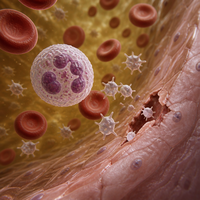
Step 2: Platelet Plug Formation
Platelets adhere to exposed collagen fibers at the injury site via von Willebrand factor. They become sticky, swell, and release ADP and serotonin to recruit more platelets, forming a temporary plug.
Step 3: Coagulation Cascade
Coagulation reinforces the platelet plug with a fibrin mesh. Two pathways initiate the cascade:
Intrinsic Pathway: Triggered by negatively charged surfaces (e.g., collagen). All factors are present in the blood. Slower but forms robust clots.
Extrinsic Pathway: Triggered by tissue factor (Factor III) from damaged tissue. Bypasses several steps, forming clots rapidly (within 15 seconds).
Both pathways require Ca2+ and PF3, converging at Factor X to enter the common pathway.
The Common Pathway
Factor X, Ca2+, PF3, and Factor V form prothrombin activator.
Prothrombin activator converts prothrombin to thrombin.
Thrombin converts fibrinogen to fibrin, forming the stable mesh.
Thrombin also activates Factor XIII, which cross-links fibrin strands.
Clot Retraction and Fibrinolysis
After clot formation, platelets contract (using actin and myosin) to pull vessel edges together and squeeze out serum. Endothelial cells secrete tissue plasminogen activator (tPA), converting plasminogen to plasmin, which digests fibrin and dissolves the clot after repair.
Hemostatic Disorders
Clinical Applications
Thrombus: Clot persisting in an unbroken vessel.
Embolus: A thrombus that breaks free and travels in the bloodstream.
Thrombocytopenia: Platelet count <50,000/µL, causing spontaneous bleeding.
Hemophilia: Hereditary deficiency of clotting factors (e.g., Factor VIII).
Blood Typing: ABO and Rh Systems
ABO Blood Grouping
Blood types are defined by the presence or absence of A and B antigens on RBCs and corresponding antibodies in plasma:
Blood Type | RBC Antigens | Plasma Antibodies |
|---|---|---|
A | A | Anti-B |
B | B | Anti-A |
AB | A, B | None |
O | None | Anti-A, Anti-B |
The Rh Blood Group
The Rh system is based on the presence (Rh+) or absence (Rh-) of the D antigen. Unlike ABO antibodies, anti-Rh antibodies are not preformed and only develop after exposure to Rh+ blood. Hemolytic disease of the newborn can occur if an Rh- mother is sensitized and carries an Rh+ fetus.
Transfusion Reactions
If incompatible blood is transfused, recipient antibodies bind donor antigens, causing agglutination and hemolysis. Free hemoglobin can block kidney tubules, leading to acute renal failure.
Blood Typing Laboratory
Blood typing is performed by mixing RBCs with anti-A, anti-B, and anti-D serums and observing agglutination. Type O- is the universal donor; AB+ is the universal recipient.
Test Serum | Agglutination? | Interpretation |
|---|---|---|
Anti-A | Yes | A antigen present |
Anti-B | Yes | B antigen present |
Anti-D | Yes | Rh positive |
Summary Table: Formed Elements of Blood
Element | Main Types | Function |
|---|---|---|
Leukocytes | Granulocytes (neutrophils, eosinophils, basophils); Agranulocytes (lymphocytes, monocytes) | Defense against pathogens |
Thrombocytes | Platelets | Hemostasis (clotting) |
Erythrocytes | Red blood cells | Oxygen and CO2 transport |
Key Takeaways
Leukocytes defend against infection; platelets mediate clotting; blood typing ensures transfusion safety.
Hemostasis involves vascular spasm, platelet plug formation, and coagulation.
ABO and Rh antigens determine compatibility; transfusion reactions can be life-threatening.
