 뒤로
뒤로Membrane Dynamics and Transport Mechanisms in Human Physiology
스터디 가이드 - 스마트 노트
 자료에 맞춘 맞춤형 노트, 핵심 정의, 예시, 맥락을 확장해 제공합니다.
Body Fluid Compartments
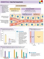
Overview of Body Fluid Compartments
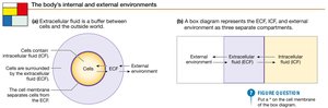
The human body is composed of various fluid compartments that are essential for physiological processes. These compartments are separated by selectively permeable membranes, allowing for the regulation of solute and water movement.
Intracellular Fluid (ICF): Fluid contained within cells; makes up about two-thirds of total body water.
Extracellular Fluid (ECF): Fluid outside cells, including interstitial fluid (between cells) and plasma (within blood vessels).
Exchange: Movement of water and solutes between compartments is tightly regulated to maintain homeostasis.
Membrane Permeability and Homeostasis
Selective Permeability and Solute Types
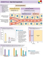
Cell membranes are selectively permeable, allowing some substances to cross more easily than others. This property is crucial for maintaining chemical and electrical gradients across the membrane.
Penetrating (permeable) solutes: Can cross the membrane freely (e.g., urea).
Non-penetrating (impermeable) solutes: Cannot cross the membrane (e.g., Na+, Cl-).
Chemical disequilibrium: Unequal distribution of solutes across compartments.
Electrical disequilibrium: Unequal distribution of charges, leading to membrane potential.
Homeostasis ≠ Equilibrium: Homeostasis maintains a dynamic steady state, not true equilibrium.
Osmosis and Osmotic Pressure
Principles of Osmosis
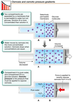
Osmosis is the movement of water across a selectively permeable membrane, driven by differences in solute concentration. Water moves from areas of higher water concentration (lower solute concentration) to areas of lower water concentration (higher solute concentration).
Osmotic equilibrium: Achieved when water movement balances solute concentrations, typically around 300 mOsM in human cells.
Osmotic pressure: The pressure required to prevent water movement across the membrane, measured in atm or mm Hg.
Osmolarity vs. Tonicity
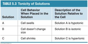
Osmolarity refers to the total concentration of solute particles in a solution, while tonicity describes how a solution affects cell volume, depending on the concentration of non-penetrating solutes.
Isotonic solution: No net change in cell volume.
Hypertonic solution: Cell shrinks as water leaves the cell.
Hypotonic solution: Cell swells as water enters the cell.
Solution | Cell Behavior When Placed in the Solution | Description of the Solution Relative to the Cell |
|---|---|---|
A | Cell swells | Solution A is hypotonic |
B | Cell doesn’t change size | Solution B is isotonic |
C | Cell shrinks | Solution C is hypertonic |
Transport Processes Across Membranes
Simple Diffusion
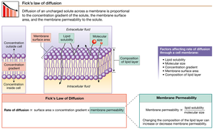
Simple diffusion is the passive movement of molecules from an area of high concentration to an area of low concentration, driven by the concentration gradient. It does not require energy input and is most effective for small, non-polar molecules such as O2 and CO2.
Fick’s Law of Diffusion: The rate of diffusion is proportional to surface area, concentration gradient, and membrane permeability.
Protein-Mediated Transport
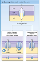
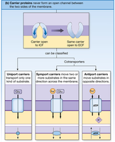
Transport proteins facilitate the movement of specific molecules across the cell membrane. These include channel proteins and carrier proteins.
Channel proteins: Create water-filled pores for ions or water to pass through. Can be open (leak channels) or gated (ligand, voltage, or mechanically gated).
Carrier proteins: Bind to substrates and undergo conformational changes to transport molecules. Types include uniport (one substance), symport (two or more substances in the same direction), and antiport (substances in opposite directions).
Facilitated Diffusion
Facilitated diffusion is a passive process where carrier proteins transport molecules down their concentration gradient without energy input. An example is the GLUT transporter for glucose.
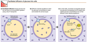
Active Transport
Active transport moves substances against their concentration gradients and requires energy, usually from ATP.
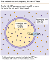
Primary active transport: Direct use of ATP, e.g., Na+/K+ ATPase pump, which moves 3 Na+ out and 2 K+ into the cell per ATP hydrolyzed.
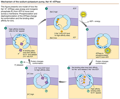
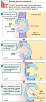
Secondary active transport: Indirect use of ATP; uses the energy stored in ion gradients created by primary active transport. Example: Sodium-glucose transporter (SGLT).
Transporter Protein Properties
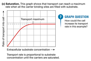
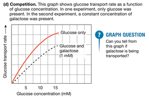
Transport proteins exhibit specificity (bind only certain molecules), competition (similar molecules compete for transport), and saturation (transport rate plateaus when all carriers are occupied).
Summary of Transport Mechanisms
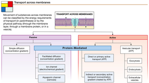
Transport across membranes can be classified as passive (simple diffusion, facilitated diffusion, ion channels, aquaporins) or active (primary/secondary active transport, vesicular transport).
Vesicular Transport
Types of Vesicular Transport
Vesicular transport involves the movement of large particles or volumes of fluid via vesicles. Types include:
Endocytosis: Uptake of material into the cell by forming vesicles.
Phagocytosis: "Cell eating"; uptake of large particles.
Pinocytosis: "Cell drinking"; uptake of extracellular fluid.
Exocytosis: Release of material from the cell by vesicle fusion with the membrane.
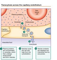
Transcytosis: Movement of substances across a cell via endocytosis and exocytosis.
Epithelial Transport
Structure and Function of Epithelia
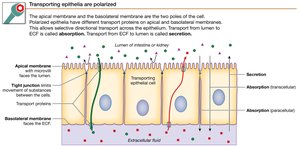
Epithelial cells line surfaces and regulate the movement of substances between the internal and external environments. They have distinct apical (facing lumen) and basolateral (facing interstitial fluid) surfaces.
Transcellular pathway: Movement through the cell.
Paracellular pathway: Movement between cells.
Absorption: Movement from lumen to ECF.
Secretion: Movement from ECF to lumen.
Transcellular Absorption of Glucose
Glucose absorption in the intestine or kidney involves coordinated action of multiple transporters:
SGLT (Na+-glucose symporter): Located on the apical membrane; uses Na+ gradient to transport glucose into the cell (secondary active transport).
GLUT transporter: Located on the basolateral membrane; facilitates glucose diffusion into the ECF (passive transport).
Na+-K+ ATPase: Located on the basolateral membrane; maintains Na+ gradient by pumping Na+ out and K+ in (primary active transport).
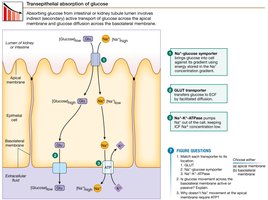 *Additional info: The coordinated function of these transporters ensures efficient absorption of glucose and maintenance of ion gradients essential for cell function.*
