 뒤로
뒤로Microscopy: Structure, Function, and Use in Anatomy & Physiology
스터디 가이드 - 스마트 노트
 자료에 맞춘 맞춤형 노트, 핵심 정의, 예시, 맥락을 확장해 제공합니다.
Lab: The Microscope
Introduction to Microscopy
The microscope is an essential tool in Anatomy & Physiology, allowing for the observation of cells, tissues, and microorganisms that are invisible to the naked eye. Understanding the structure and function of the microscope is foundational for laboratory work in biological sciences.
Microscope: An optical instrument used to magnify small objects.
Compound Microscope: Uses multiple lenses to achieve higher magnification and resolution.
Proper Handling: Always carry with two hands—one on the arm and one under the base.
Clean Lenses: Only with lens paper to avoid scratches.
Start with Lowest Power: Begin focusing with the lowest power objective to locate specimens easily.
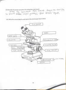
Parts of the Compound Microscope
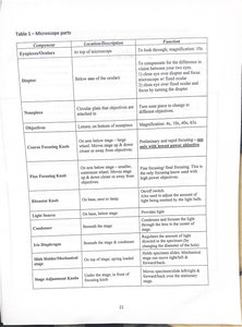
Each part of the microscope has a specific function that contributes to the magnification and clarity of the specimen being observed.
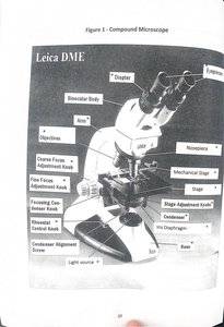
Component | Location/Description | Function |
|---|---|---|
Eyepiece/Oculars | At top of microscope | To look through; magnifies 10x |
Diopter | Below one of the oculars | Compensates for differences in vision between eyes |
Nosepiece | Circular plate that objectives are attached to | Turns to change objective lens |
Objectives | Lower, on bottom of nosepiece | Magnification: 4x, 10x, 40x, 63x |
Coarse Focusing Knob | On either side | Large, rapid focusing (use only with lowest power) |
Fine Focusing Knob | On either side, smaller | Fine focusing (use with all objectives) |
Stage | Beneath objectives | Holds slide |
Stage Adjustment Knob | Below stage | Moves slide left/right & forward/back |
Light Source | Base | Illuminates specimen |
Condenser | Beneath stage | Focuses light onto specimen |
Condenser Adjustment Knob | Below stage | Raises/lowers condenser |
Iris Diaphragm | Below condenser | Regulates light intensity |
Using the Microscope: Focusing and Magnification
Proper focusing and understanding magnification are crucial for clear observation of specimens.
Start with the scanning objective (lowest power) and coarse focus.
Switch to higher objectives for greater detail, using only the fine focus knob.
Adjust the iris diaphragm and condenser for optimal lighting.
Never use the coarse focus with high power objectives to avoid damaging slides or lenses.
Calculating Total Magnification
Total magnification is the product of the ocular lens and the objective lens magnifications:
For example, with a 10x ocular and a 40x objective:
Field of View and Working Distance
The field of view is the visible area when looking through the microscope. The working distance is the space between the objective lens and the specimen.
As magnification increases, the field of view and working distance decrease.
Field of view can be measured using a ruler under the microscope at low power, then calculated for higher powers.
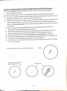
Estimating Field of View
To estimate the field of view at higher magnifications:
Estimating Object Size
Once the field of view is known, the size of objects can be estimated by comparing their length to the diameter of the field of view.
For example, if a cell spans half the field of view (200 μm), its size is approximately 100 μm.
Metric System and Unit Conversions
Microscopy requires familiarity with metric units such as millimeters (mm), micrometers (μm), and nanometers (nm).
1 mm = 1000 μm
1 μm = 0.001 mm
1 nm = 0.001 μm
Magnification, Drawing Size, and Actual Size
To determine the magnification of a drawing:
To find the actual size of a specimen:
Microscope Lab Review and Application
Proper microscope use includes safe transport, correct focusing, and cleaning procedures. Understanding the relationship between magnification, field of view, and working distance is essential for accurate observation and measurement in Anatomy & Physiology labs.
Always clean lenses with lens paper.
Carry the microscope with two hands.
Begin focusing with the lowest power objective.
Adjust light and diaphragm for best image quality.
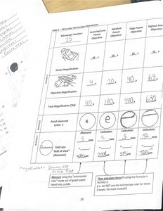
Summary Table: Objective Magnification, Field of View, and Calculations
Objective | Ocular Magnification | Objective Magnification | Total Magnification | Field of View (μm) |
|---|---|---|---|---|
Scanning | 10x | 4x | 40x | 4000 |
Low Power | 10x | 10x | 100x | 2000 |
High Power | 10x | 40x | 400x | 500 |
Oil Immersion | 10x | 100x | 1000x | 200 |
Key Terms and Concepts
Resolution: The ability to distinguish two points as separate entities.
Depth of Field: The thickness of the specimen that is in focus at one time.
Wet Mount: A slide preparation technique using a drop of liquid and a coverslip.
Oil Immersion: Technique using oil to increase resolution at high magnification.
Practical Applications
Microscopy is used to study cell structure, tissue organization, and microorganisms in Anatomy & Physiology.
Accurate measurement and observation are critical for diagnosis and research.
