 뒤로
뒤로Nervous System Structure and Resting Membrane Potential: Study Notes
스터디 가이드 - 스마트 노트
 자료에 맞춘 맞춤형 노트, 핵심 정의, 예시, 맥락을 확장해 제공합니다.
Overview of the Nervous System
Central and Peripheral Divisions
The nervous system is divided into the Central Nervous System (CNS) and the Peripheral Nervous System (PNS). The CNS consists of the brain and spinal cord, while the PNS includes all neural tissue outside the CNS. The PNS is further subdivided into afferent (sensory) and efferent (motor) divisions.
Afferent Division: Transmits sensory information from receptors to the CNS.
Efferent Division: Carries motor commands from the CNS to effectors (muscles and glands).
Somatic Nervous System: Controls voluntary movements via skeletal muscles.
Autonomic Nervous System: Regulates involuntary functions (e.g., cardiac muscle, smooth muscle, glands) and is further divided into sympathetic, parasympathetic, and enteric systems.
Cells of the Nervous System
Neurons and Glial Cells
The nervous system contains two main types of cells: neurons (excitable cells that transmit electrical signals) and glial cells (support cells that maintain the environment around neurons).
Neurons: Specialized for communication via electrical and chemical signals.
Glial Cells: Outnumber neurons and provide structural, metabolic, and functional support.
Structure of a Neuron
Neurons have a characteristic structure that supports their function in signal transmission.
Soma (Cell Body): Contains the nucleus and most organelles.
Dendrites: Receive incoming signals from other neurons.
Axon: Conducts action potentials away from the soma.
Axon Hillock: Site where action potentials are initiated.
Axon Terminals: Release neurotransmitters to communicate with other cells.
Axonal Transport
Materials are transported along the axon via microtubules and motor proteins. This process is essential for neuron function and survival.
Anterograde Transport: Moves materials from the soma to the axon terminal (e.g., neurotransmitters).
Retrograde Transport: Moves materials from the axon terminal back to the soma.
Transport Rates: Slow (0.5–40 mm/day) and fast (100–400 mm/day) transport mechanisms exist.
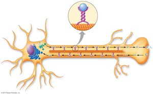
Synapse
A synapse is the site of communication between two neurons or between a neuron and an effector organ. Neurotransmitters released from the presynaptic neuron bind to receptors on the postsynaptic cell, transmitting the signal.
Ion Channels in Neurons
Neuronal membranes contain several types of ion channels that regulate membrane potential and signal transmission:
Leak Channels: Always open; contribute to resting membrane potential.
Ligand-Gated Channels: Open in response to neurotransmitter binding; found on dendrites and soma.
Voltage-Gated Channels: Open in response to changes in membrane potential; crucial for action potentials.
Calcium Channels: Located at axon terminals; trigger neurotransmitter release.
Structural and Functional Classes of Neurons
Neurons are classified based on structure (e.g., multipolar, bipolar, pseudo-unipolar) and function (afferent, efferent, interneurons).
Afferent Neurons: Carry sensory information to the CNS.
Efferent Neurons: Transmit commands from the CNS to effectors.
Interneurons: Integrate information within the CNS.
Structural Organization in Nervous Tissue
Nuclei: Clusters of neuron cell bodies in the CNS.
Ganglia: Clusters of neuron cell bodies in the PNS.
Tracts/Pathways: Bundles of axons in the CNS.
Nerves: Bundles of axons in the PNS.
Glial Cells and Myelination
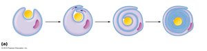
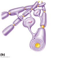
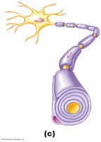
Glial cells support neurons and form myelin sheaths, which insulate axons and increase the speed of electrical transmission.
Astrocytes: Maintain the blood-brain barrier and provide metabolic support.
Microglia: Act as immune cells in the CNS.
Oligodendrocytes: Myelinate axons in the CNS; one cell can myelinate multiple axons.
Schwann Cells: Myelinate axons in the PNS; each cell myelinates a single axon segment.
Resting Membrane Potential
Establishment of Resting Membrane Potential
All cells have a resting membrane potential, which is the voltage difference across the plasma membrane when the cell is not actively sending signals. In neurons, this is typically around –70 mV, with the inside of the cell being more negative than the outside.
Key Factors: Ion concentration gradients and membrane permeability to specific ions.
Typical Ion Distribution: High Na+ and Cl– outside; high K+ and organic anions inside.
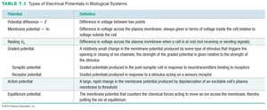
Types of Electrical Potentials in Biological Systems
The table below summarizes the main types of electrical potentials relevant to neurons:
Potential | Definition |
|---|---|
Potential difference (V) | Difference in voltage between two points |
Membrane potential (Vm) | Difference in voltage across the plasma membrane (inside relative to outside) |
Resting potential | Membrane voltage when a cell is not sending signals |
Graded potential | Change in membrane potential with varying strength, usually short distance |
Synaptic potential | Graded potential at a synapse in response to neurotransmitter binding |
Receptor potential | Graded potential in response to a stimulus acting on a sensory receptor |
Action potential | Large, rapid change in membrane potential due to depolarization and repolarization |
Equilibrium potential | Membrane potential at which the chemical and electrical forces for an ion are balanced |
Role of the Na+/K+ Pump
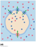
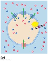
The Na+/K+-ATPase (sodium-potassium pump) is essential for maintaining the resting membrane potential. It actively transports 3 Na+ ions out of the cell and 2 K+ ions into the cell, creating and maintaining the necessary ion gradients.
Direct Contribution: Electrogenic effect (net +1 charge out per cycle).
Indirect Contribution: Maintains concentration gradients for Na+ and K+.
Equilibrium Potentials
The equilibrium potential for an ion is the membrane voltage at which the electrical and chemical driving forces for that ion are equal and opposite. It can be calculated using the Nernst equation:
Potassium (K+):
Sodium (Na+):
The Nernst equation (for a monovalent cation):
where is the gas constant, is temperature in Kelvin, is the ion charge, and is Faraday's constant.
Resting Membrane Potential in Neurons
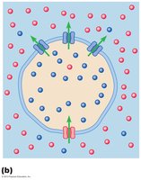
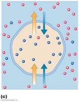
Neurons are more permeable to K+ than Na+ (about 25 times more), so the resting membrane potential is closer to the K+ equilibrium potential. The steady state is achieved when the inflow of Na+ is balanced by the outflow of K+, resulting in a stable resting potential of approximately –70 mV.
Summary Table: Key Ion Movements and Forces
K+: Chemical force drives out; electrical force pulls in; equilibrium at –94 mV.
Na+: Chemical force drives in; electrical force pushes out; equilibrium at +60 mV.
Resting Potential: More K+ leaves than Na+ enters, making the inside negative.
Electrical Signals in Neurons
Generation of Electrical Signals
Changes in membrane potential occur due to the opening and closing of gated ion channels in response to stimuli. These changes underlie the generation and propagation of electrical signals in neurons, including graded potentials and action potentials.
