 뒤로
뒤로Stem Cells and Body Membranes: Structure, Types, and Functions
스터디 가이드 - 스마트 노트
 자료에 맞춘 맞춤형 노트, 핵심 정의, 예시, 맥락을 확장해 제공합니다.
Stem Cells
Characteristics of Stem Cells
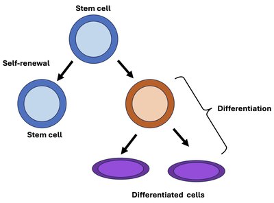
Stem cells are unique cells in the body with the ability to both self-renew and differentiate into specialized cell types. These properties are essential for growth, development, and tissue repair.
Self-renewal: The capacity to divide and produce more stem cells, maintaining the stem cell pool throughout life.
Potency: The ability to differentiate into different cell types. Stem cells are classified by their potency:
Totipotent: Can give rise to all cell types, including embryonic and extraembryonic tissues.
Pluripotent: Can differentiate into nearly all cell types derived from the three germ layers (ectoderm, mesoderm, endoderm).
Multipotent: Can develop into a limited range of cell types within a particular tissue or organ.
Unipotent: Can produce only one cell type, but retain the property of self-renewal.
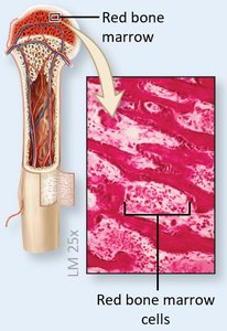
Example: Hematopoietic stem cells in bone marrow are multipotent, giving rise to various blood cells.
Embryonic Stem Cells
Types and Developmental Stages
Embryonic stem cells are derived from early-stage embryos and are highly potent, playing a critical role in development.
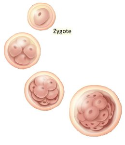
Totipotent Stem Cells: Present in the zygote and early cleavage stages; can form all embryonic and extraembryonic tissues.
Pluripotent Stem Cells: Found in the inner cell mass (embryoblast) of the blastocyst; can form all tissues of the body but not extraembryonic tissues.
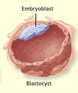
Example: Pluripotent stem cells from the blastocyst can be cultured in the lab to study development or for regenerative medicine.
Adult Stem Cells
Types and Locations
Adult stem cells are found in various tissues after development and are primarily responsible for tissue maintenance and repair.
Multipotent Stem Cells: Can differentiate into a limited range of cells within a specific tissue (e.g., hematopoietic stem cells in bone marrow).
Unipotent Stem Cells: Can produce only one cell type but are important for tissue regeneration (e.g., epithelial stem cells in the skin).
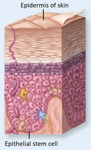
Example: Epithelial stem cells in the skin continually replace cells lost from the surface.
Body Membranes
Overview and Classification
Body membranes are thin sheets of tissue that cover body surfaces, line cavities, and form protective barriers. They are classified based on location and structure.
Internal membranes: Mucous, serous, and synovial membranes.
External membrane: Cutaneous membrane (skin).
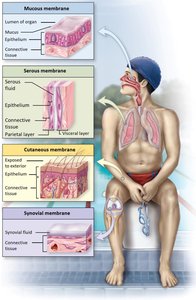
Serous Membranes
Structure and Function
Serous membranes line body cavities that do not open to the exterior and secrete serous fluid to reduce friction between organs.
Two layers:
Parietal layer: Lines the cavity wall.
Visceral layer: Covers the organs within the cavity.
Thoracic Cavity
Pleural Cavity
Parietal pleura
Visceral pleura = organ surface
Pericardial Cavity
Parietal pericardium
Visceral pericardium = organ surface
Abdominopelvic Cavity
Peritoneum
Parietal peritoneum
Visceral peritoneum = organ surface
Examples: Pleura (lungs), pericardium (heart), peritoneum (abdominopelvic organs).
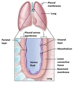
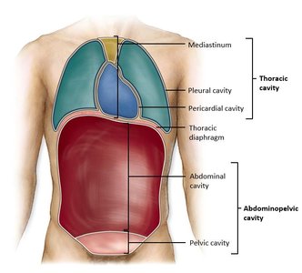
Example: The pleural membranes surround the lungs and secrete serous fluid to allow smooth lung movement during breathing.
Mucous Membranes
Structure and Function
Mucous membranes line body cavities that are open to the external environment, such as the digestive, respiratory, urinary, and reproductive tracts. They secrete mucus to protect and lubricate surfaces.
Components: Columnar epithelium with goblet cells, below is areolar connective tissue (lamina propria)
Lines: digestive tract, urinary tract, respiratory tract, reproductive tract
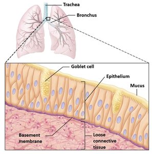
Example: The mucous membrane of the respiratory tract traps dust and microbes, aiding in their removal.
Synovial Membranes
Structure and Function
Synovial membranes line the cavities of freely movable joints (synovial joints) and secrete synovial fluid for lubrication and nourishment of joint tissues.
Components: simple squamous epithelium, areolar connective tissue
Synovial Fluid: in the joint cavity that helps reduce friction
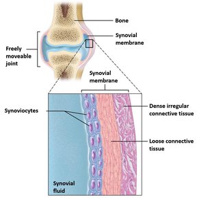
Example: The knee joint is lined by a synovial membrane that produces fluid to reduce friction during movement.
Cutaneous Membrane
Structure and Function
The cutaneous membrane, or skin, is the body's largest organ and serves as a protective barrier against environmental hazards. It consists of two main layers:
Epidermis: Keratinized stratified squamous epithelium that provides waterproofing and protection.
Dermis: Dense irregular connective tissue that supports and nourishes the epidermis.
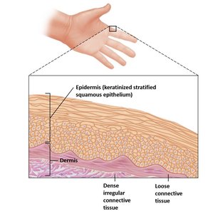
Example: The skin protects underlying tissues from mechanical damage, pathogens, and dehydration.
Membrane Type | Location | Main Function |
|---|---|---|
Mucous | Lines cavities open to exterior (e.g., respiratory, digestive tracts) | Protection, secretion of mucus |
Serous | Lines closed body cavities (e.g., pleura, pericardium, peritoneum) | Reduces friction via serous fluid |
Synovial | Lines joint cavities | Lubrication, nourishment of joint tissues |
Cutaneous | External body surface (skin) | Protection, sensation, thermoregulation |
