 뒤로
뒤로The Cell: Anatomy and Division – Study Notes
스터디 가이드 - 스마트 노트
 자료에 맞춘 맞춤형 노트, 핵심 정의, 예시, 맥락을 확장해 제공합니다.
The Cell: Anatomy and Division
Introduction to the Cell
The cell is the fundamental structural and functional unit of all living organisms. Human cells display a wide variety of shapes, sizes, and internal structures, reflecting their specialized roles. Despite this diversity, all cells share certain basic anatomical features and functions necessary for life, such as maintaining boundaries, metabolizing, digesting nutrients, disposing of wastes, growing, reproducing, moving, and responding to stimuli.
Anatomy of the Composite (Generalized) Cell
Major Regions of the Cell
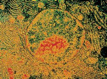
Nucleus: Usually located near the center, contains genetic material (DNA) and controls cellular activities.
Cytoplasm: The region between the nucleus and plasma membrane, containing organelles suspended in cytosol.
Plasma Membrane: The outer boundary that separates the cell from its environment and regulates the movement of substances in and out.
Nucleus
Genetic Material: DNA is organized as chromatin when the cell is not dividing and condenses into chromosomes during cell division.
Nucleolus: Site of ribosome assembly, composed of proteins and RNA.
Nuclear Envelope: Double-layered membrane with nuclear pores allowing passage of proteins and RNA.
Plasma Membrane
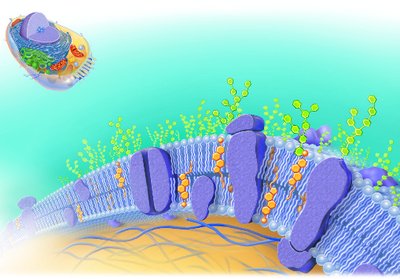
The plasma membrane is a selectively permeable barrier composed mainly of a phospholipid bilayer with embedded proteins, cholesterol, and carbohydrate side chains. It regulates the entry and exit of substances, maintains cellular integrity, and facilitates cell signaling and interactions.
Selective Permeability: Allows certain molecules to pass while restricting others.
Transport Mechanisms: Includes active (requires ATP) and passive (driven by concentration/pressure gradients) transport.
Microvilli: Fingerlike projections that increase surface area for absorption.
Cytoplasm and Organelles
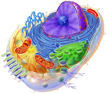
The cytoplasm is the site of most cellular activities and contains various organelles, each with specialized functions:
Ribosomes: Sites of protein synthesis; found free in cytoplasm or attached to rough ER.
Endoplasmic Reticulum (ER): Network of membranous tubules; rough ER (with ribosomes) synthesizes proteins, smooth ER synthesizes lipids and detoxifies chemicals.
Golgi Apparatus: Modifies, sorts, and packages proteins and lipids for secretion or delivery to other organelles.
Lysosomes: Contain digestive enzymes for breaking down waste and cellular debris.
Peroxisomes: Detoxify harmful substances using oxidase enzymes.
Mitochondria: "Powerhouses" of the cell, generate ATP through oxidative respiration.
Cytoskeleton: Network of microfilaments, intermediate filaments, and microtubules providing structural support and facilitating movement.
Centrioles: Organize the mitotic spindle during cell division and form bases of cilia and flagella.
Inclusions: Non-living materials such as glycogen granules, lipid droplets, and pigments.
Summary Table: Structure and Function of Cytoplasmic Organelles
Organelle | Location and Function |
|---|---|
Ribosomes | Sites of protein synthesis; free or attached to rough ER |
Endoplasmic Reticulum (ER) | Rough ER: protein synthesis and transport; Smooth ER: lipid synthesis, metabolism, detoxification |
Golgi Apparatus | Packages proteins/lipids for export or incorporation into membranes; forms lysosomes |
Lysosomes | Digest worn-out organelles and foreign substances |
Peroxisomes | Detoxify alcohol, hydrogen peroxide, and other chemicals |
Mitochondria | Produce ATP via oxidative metabolism |
Centrioles | Direct mitotic spindle formation; form bases of cilia/flagella |
Cytoskeletal Elements | Support cell structure, intracellular transport, cell movement |
Cell Diversity and Specialization
Comparing Cell Types
Cells differ in shape, size, and presence of specialized structures, reflecting their functions:
Simple Squamous Epithelium: Flat cells for diffusion/filtration.
Smooth Muscle Cells: Elongated for contraction and movement.
Red Blood Cells: Biconcave, anucleate for efficient gas transport.
Sperm Cells: Flagellated for motility.
Cell shape is closely related to function; for example, microvilli increase absorption, while flagella enable movement.
Cell Division: Mitosis and Cytokinesis
Overview of the Cell Cycle
The cell cycle consists of interphase (growth and normal activities) and cell division (mitosis and cytokinesis). Mitosis ensures genetic continuity by producing two genetically identical daughter cells.
Stages of Mitosis
Interphase: Cell grows, DNA replicates, centrioles replicate.
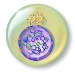
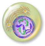
Prophase: Chromatin condenses into chromosomes; nuclear envelope breaks down; spindle forms.
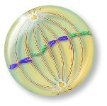
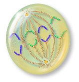
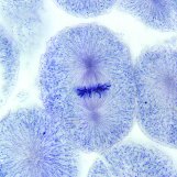
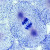
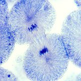
Metaphase: Chromosomes align at the metaphase plate (cell equator).
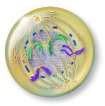
Anaphase: Sister chromatids separate and move toward opposite poles.
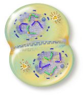
Telophase: Chromosomes decondense; nuclear envelopes reform; spindle disappears.
Cytokinesis: Cytoplasm divides, forming two daughter cells.
Microscopic Identification of Mitotic Stages
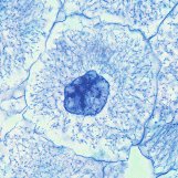
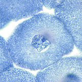
Mitotic stages can be observed in rapidly dividing tissues, such as the whitefish blastula. Each stage has distinct morphological features under the microscope.
Importance of Mitosis
Ensures genetic continuity during growth and tissue repair.
Maintains chromosome number in somatic cells.
Distinguished from meiosis, which produces genetically diverse gametes.
Key Terms and Definitions
Cell: The basic unit of structure and function in living organisms.
Organelle: Specialized subunit within a cell with a specific function.
Inclusion: Non-living material in the cytoplasm (e.g., glycogen, lipid droplets).
Interphase: Period of cell growth and DNA replication between divisions.
Mitosis: Division of the nucleus to produce two identical nuclei.
Cytokinesis: Division of the cytoplasm, resulting in two daughter cells.
Summary Table: Phases of Mitosis and Key Events
Phase | Key Events |
|---|---|
Prophase | Chromatin condenses, spindle forms, nuclear envelope breaks down |
Metaphase | Chromosomes align at metaphase plate |
Anaphase | Sister chromatids separate and move to poles |
Telophase | Chromosomes decondense, nuclear envelope reforms |
Cytokinesis | Cytoplasm divides, forming two cells |
Additional info:
Cells such as skeletal muscle, cardiac muscle, and mature neurons rarely divide after development, making injuries to these tissues particularly serious.
Red blood cells are anucleate at maturity, maximizing space for hemoglobin and gas transport, but they originate from nucleated precursors in the bone marrow.
