 뒤로
뒤로The Cell and Plasma Membrane: Structure and Transport Mechanisms
스터디 가이드 - 스마트 노트
 자료에 맞춘 맞춤형 노트, 핵심 정의, 예시, 맥락을 확장해 제공합니다.
The Cell: Structure and Diversity
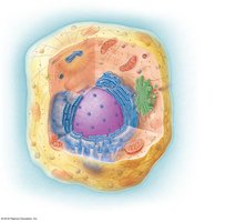
Overview of Cell Structure
Cells are the fundamental units of life, each surrounded by a plasma membrane and containing cytoplasm and a nucleus. The cytoplasm includes cytosol, organelles, and the cytoskeleton, all of which contribute to cellular function and structure.
Plasma membrane: Acts as a selective barrier between the cell's internal environment and the extracellular fluid.
Cytoplasm: The region between the plasma membrane and the nucleus, containing cytosol (fluid), organelles, and the cytoskeleton.
Nucleus: Contains genetic material (DNA) and controls cellular activities.
Types of Cells
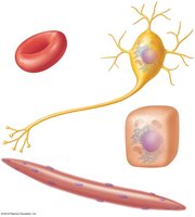
Cells in the human body vary greatly in shape and function, reflecting their specialized roles.
Red blood cells: Specialized for oxygen transport.
Nerve cells (neurons): Specialized for communication and signal transmission.
Skeletal muscle cells: Specialized for contraction and movement.
Epithelial cells: Form protective layers and are involved in absorption and secretion.
Fluid Compartments in the Body
Intracellular and Extracellular Fluid
The body's water is distributed between two main compartments:
Intracellular fluid (ICF): Fluid inside cells.
Extracellular fluid (ECF): Fluid outside cells, further divided into:
Interstitial fluid: Surrounds cells.
Plasma: Fluid component of blood.
The Plasma Membrane: Structure and Function
Fluid Mosaic Model
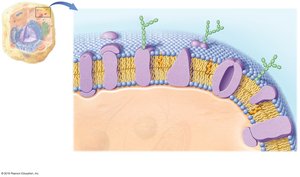
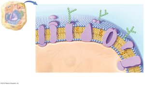
The plasma membrane is described by the fluid mosaic model, which highlights its dynamic nature and diverse components. It acts as a boundary between the ECF and ICF, controlling the movement of substances into and out of the cell.
Phospholipid bilayer: Forms the basic structure, with hydrophilic (polar) heads facing outward and hydrophobic (nonpolar) tails facing inward.
Proteins: Integral (transmembrane) and peripheral proteins serve various functions such as transport, signaling, and support.
Carbohydrates: Glycoproteins and glycolipids are involved in cell recognition and signaling.
Cholesterol: Maintains membrane fluidity and stability.
Phospholipid Structure and Bilayer Formation
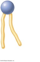
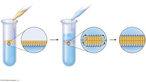
Phospholipids are amphipathic molecules with a hydrophilic head and two hydrophobic tails. In water, they spontaneously form bilayers, which are the foundation of cellular membranes.
Polar (hydrophilic) head: Attracted to water.
Nonpolar (hydrophobic) tails: Repelled by water, face inward.
Selective Permeability of the Plasma Membrane
The plasma membrane is selectively permeable, allowing some molecules to cross freely while restricting others.
Freely permeable: Non-polar, lipid-soluble, small molecules.
Require transport proteins: Most polar, water-soluble, large, or charged molecules (ions).
Functions of Membrane Proteins
Membrane proteins are essential for various cellular processes:
Transporters: Channels and carriers move substances across the membrane.
Receptors: Bind ligands to trigger cellular responses.
Enzymes: Catalyze chemical reactions at the membrane surface.
Structural support: Anchor the membrane to the cytoskeleton or extracellular matrix.
Linkers: Connect adjacent cells.
Membrane Transport Mechanisms
Overview of Membrane Transport
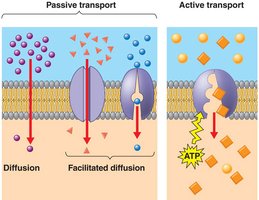
Transport across the plasma membrane can be passive (no energy required) or active (energy required).
Passive transport: Simple diffusion, facilitated diffusion, osmosis.
Active transport: Primary active transport, secondary active transport, vesicular transport (endocytosis and exocytosis).
Passive Transport
Simple Diffusion
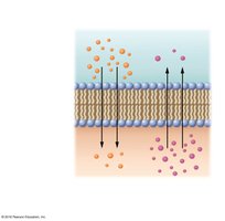
Movement of solutes from an area of high concentration to low concentration, down their concentration gradient. Nonpolar, uncharged molecules (e.g., O2, CO2) cross the membrane by simple diffusion.
Facilitated Diffusion
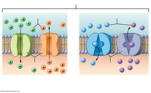
Polar and charged solutes move across the membrane with the help of protein transporters (channels or carriers). This process does not require energy and moves substances down their concentration gradient.
Channels: Create a tunnel for ions or water to pass through.
Carriers: Bind the solute and change shape to transport it across the membrane.
Osmosis
Osmosis is the diffusion of water across a selectively permeable membrane from an area of high water concentration (low solute) to low water concentration (high solute).
Osmolarity: Number of solute particles per liter of solution.
Osmotic pressure: Pulling force exerted by non-penetrating solute particles.
Tonicity: The effect of a solution on cell volume (isotonic, hypertonic, hypotonic).
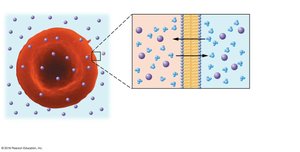
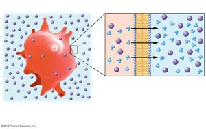
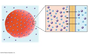
Tonicity and Red Blood Cells
Isotonic solution: Equal solute concentration inside and outside the cell; no net water movement.
Hypertonic solution: Higher solute concentration outside the cell; water leaves the cell, causing it to shrink (crenate).
Hypotonic solution: Lower solute concentration outside the cell; water enters the cell, causing it to swell and possibly burst (lyse).
Active Transport
Active transport requires energy (usually ATP) to move substances against their concentration gradients.
Primary active transport: Direct use of ATP to transport molecules (e.g., Na+/K+ pump).
Secondary active transport: Uses the energy stored in ion gradients created by primary active transport to move other substances.
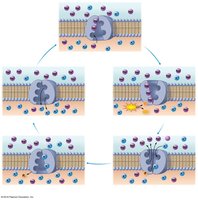
Na+/K+ Pump (Primary Active Transport)
This pump moves 3 Na+ ions out of the cell and 2 K+ ions into the cell, maintaining essential ion gradients for cell function.
Antiport mechanism: Moves ions in opposite directions.
Requires ATP hydrolysis: Energy is used to change the shape of the pump and move ions against their gradients.
Secondary Active Transport
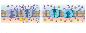
Uses the energy from the Na+ gradient (created by the Na+/K+ pump) to transport other molecules, such as glucose, into the cell.
Symport: Both substances move in the same direction.
Antiport: Substances move in opposite directions.
Vesicular Transport
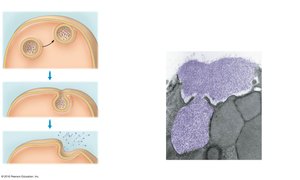
Vesicular transport moves large particles or volumes of fluid into or out of the cell using vesicles.
Endocytosis: Brings substances into the cell (includes phagocytosis and pinocytosis).
Exocytosis: Releases substances from the cell.
Summary Table: Plasma Membrane Transport
Type of Transport | Definition | Example(s) |
|---|---|---|
Simple Diffusion | Movement of solute down its concentration gradient through the phospholipid bilayer without energy input | Oxygen, Carbon dioxide, Lipids |
Facilitated Diffusion | Movement of solute down its concentration gradient with the help of membrane proteins (channels or carriers) | Ions, Glucose, Amino acids |
Osmosis | Movement of water across a selectively permeable membrane from low to high solute concentration | Water balance in cells |
Primary Active Transport | Movement of solute against its concentration gradient using ATP directly | Na+/K+ pump |
Secondary Active Transport | Movement of solute with the help of energy stored in ion gradients created by primary active transport | Glucose/Na+ symport |
Vesicular Transport | Movement of large particles or fluids via vesicles | Phagocytosis, Pinocytosis, Exocytosis |
Resting Membrane Potential
Establishment of Membrane Potential
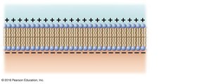
As ions move across the plasma membrane, an uneven distribution of charges is created. The inside of the cell becomes more negative compared to the outside, resulting in a resting membrane potential of approximately -70 mV. This electrical potential is essential for the function of excitable cells such as neurons and muscle cells.
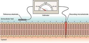
Additional info: The resting membrane potential is maintained by the selective permeability of the membrane to ions and the activity of the Na+/K+ pump. It is crucial for the generation of action potentials in nerve and muscle cells.
