 뒤로
뒤로The Cell: Structure, Function, and Life Cycle (Anatomy & Physiology I, Chapter 2)
스터디 가이드 - 스마트 노트
 자료에 맞춘 맞춤형 노트, 핵심 정의, 예시, 맥락을 확장해 제공합니다.
The Cell
Overview of Cells
Cells are the fundamental structural and functional units of all living organisms. According to cell theory, all living things are composed of cells, which arise only from pre-existing cells. Human bodies contain approximately 50-100 trillion cells, each specialized for particular functions.
Cell Theory: The cell is the smallest unit of life; all organisms are made of one or more cells; cells arise from other cells.
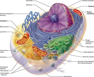
Generalized Cell Structure: All human cells share three main components:
Plasma membrane: Separates the intracellular and extracellular environments.
Cytoplasm: Contains organelles and cytosol.
Nucleus: Contains genetic material (DNA).
Plasma Membrane
Structure and Function
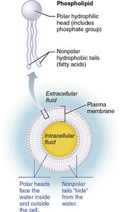
The plasma membrane is a dynamic barrier that regulates the movement of substances into and out of the cell. It is composed primarily of a phospholipid bilayer with embedded proteins and cholesterol, described by the fluid mosaic model.
Phospholipid Bilayer: Hydrophilic (water-attracting) heads face outward; hydrophobic (water-repelling) tails face inward.
Cholesterol: Increases membrane stability and fluidity.
Membrane Proteins: Integral (span the membrane) and peripheral (attached to membrane surface) proteins serve as transporters, receptors, enzymes, and cell adhesion molecules.
Membrane Specializations
Glycocalyx: Carbohydrate-rich area on cell surface for cell recognition and immune response.
Membrane Junctions:
Tight junctions: Prevent passage of substances between cells (e.g., GI tract lining).
Desmosomes: Anchor cells together, providing mechanical strength (e.g., skin, heart muscle).
Gap junctions: Allow communication and passage of ions/small molecules between cells.
Membrane Transport
The plasma membrane is selectively permeable, allowing some substances to cross more easily than others. Transport mechanisms are classified as passive (no energy required) or active (requires ATP).
Passive Transport:
Simple diffusion: Movement of nonpolar, lipid-soluble substances (e.g., O2, CO2).
Facilitated diffusion: Movement via carrier or channel proteins (e.g., glucose, ions).
Osmosis: Diffusion of water through aquaporins or directly across the bilayer.
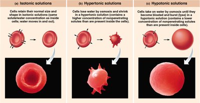
Tonicity: The effect of solution concentration on cell volume:
Isotonic: No net water movement; cell volume unchanged.
Hypertonic: Water leaves cell; cell shrinks (crenation).
Hypotonic: Water enters cell; cell swells and may burst (lysis).
Active Transport:
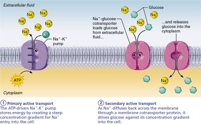
Primary active transport: Direct use of ATP (e.g., Na+-K+ pump moves 3 Na+ out and 2 K+ in per ATP hydrolyzed).
Secondary active transport: Uses energy stored in gradients created by primary active transport (e.g., Na+-glucose cotransporter).
Vesicular Transport: Movement of large particles or volumes via vesicles (requires ATP):
Endocytosis: Phagocytosis (cell eating), pinocytosis (cell drinking), receptor-mediated endocytosis.
Exocytosis: Ejection of substances (e.g., hormones, neurotransmitters).
Transcytosis: Transport into, across, and out of cell.
Vesicular trafficking: Movement within the cell.
Cell-Environment Interactions
Cell Adhesion Molecules (CAMs): Anchor cells, assist movement, attract immune cells, and transmit signals.
Plasma Membrane Receptors: Bind chemical signals (ligands) for contact or chemical signaling (e.g., G protein-coupled receptors).
Cytoplasm and Organelles
Cytoplasm
The cytoplasm is the cellular material between the plasma membrane and nucleus, consisting of cytosol (fluid), inclusions (stored nutrients/pigments), and organelles (specialized structures).
Mitochondria: Site of ATP production via cellular respiration; contains its own DNA and ribosomes.
Ribosomes: Sites of protein synthesis; free (cytosol) or membrane-bound (rough ER).
Endoplasmic Reticulum (ER):
Rough ER: Studded with ribosomes; synthesizes proteins and membranes.
Smooth ER: Lipid metabolism, detoxification, calcium storage.
Golgi Apparatus: Modifies, sorts, and packages proteins/lipids for secretion or delivery to organelles.
Lysosomes: Contain digestive enzymes for breakdown of waste, pathogens, and cellular debris.
Peroxisomes: Detoxify harmful substances; neutralize free radicals via oxidase and catalase enzymes.
Cytoskeleton: Network of protein filaments for cell shape, movement, and organelle positioning.
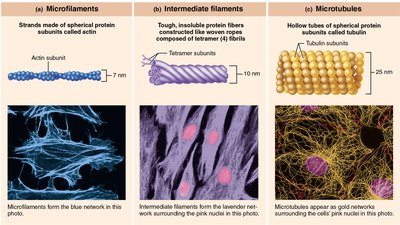
Centrosome and Centrioles: Organize microtubules; essential for cell division (mitotic spindle formation).
Surface Extensions:
Cilia: Move substances across cell surfaces (e.g., respiratory tract).
Flagella: Propel cells (e.g., sperm).
Microvilli: Increase surface area for absorption (e.g., intestines).
Nucleus
Structure and Function
The nucleus is the largest organelle, containing the genetic blueprint (DNA) for protein synthesis. It is surrounded by a double-membrane nuclear envelope with nuclear pores for molecular exchange.
Nucleolus: Site of ribosomal RNA synthesis and ribosome assembly.
Chromatin: DNA wrapped around histone proteins; condenses into chromosomes during cell division.
Chromosomes: Condensed chromatin, visible during mitosis/meiosis.
Protein Synthesis
Central Dogma of Biology
Genetic information flows from DNA to RNA to protein. This process involves two main steps: transcription and translation.
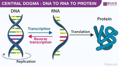
Transcription: DNA is transcribed into messenger RNA (mRNA) in the nucleus. Introns are removed, and exons are spliced together to form mature mRNA.
Translation: mRNA is translated into a polypeptide chain at the ribosome, with transfer RNA (tRNA) bringing specific amino acids according to the codon sequence.
Genetic Code: Triplet codons on mRNA specify amino acids; the code is redundant but not ambiguous.
Cell Life Cycle
Interphase and Cell Division
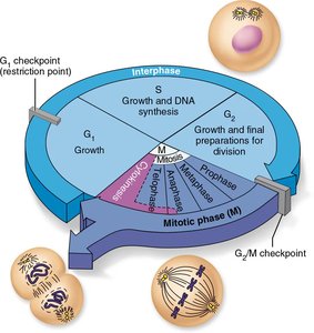
The cell cycle consists of interphase (growth and DNA replication) and the mitotic phase (cell division). Interphase includes G1 (growth), S (DNA synthesis), and G2 (preparation for division).
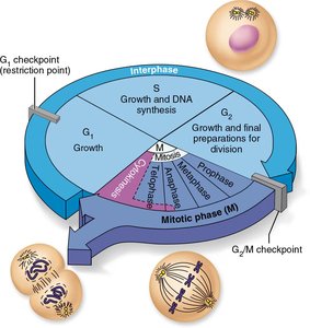
DNA Replication: Occurs during S phase; semi-conservative (each new DNA has one old and one new strand).
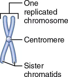
Chromosomes: After replication, each consists of two sister chromatids joined at a centromere.
Mitotic Phase (Mitosis and Cytokinesis)
Prophase: Chromatin condenses into chromosomes; nuclear envelope dissolves; spindle forms.
Metaphase: Chromosomes align at the cell equator (metaphase plate).
Anaphase: Sister chromatids separate and move to opposite poles.
Telophase: Chromosomes decondense; nuclear envelope reforms.
Cytokinesis: Cytoplasm divides, producing two genetically identical daughter cells.
Cell Specialization, Differentiation, and Aging
Cell Specialization: Cells develop unique structures and functions (e.g., neurons, RBCs, sperm).
Cell Differentiation: Stem cells become specialized cell types.
Apoptosis: Programmed cell death removes damaged or unnecessary cells.
Hyperplasia: Increased cell production.
Hypertrophy: Increased cell size.
Atrophy: Decreased cell size due to loss of stimulation or disease.
Cell Aging: Theories include wear-and-tear, immune system decline, and genetic programming (telomere shortening).
