 뒤로
뒤로The Cell: Structure, Function, and Processes in Human Anatomy & Physiology
스터디 가이드 - 스마트 노트
 자료에 맞춘 맞춤형 노트, 핵심 정의, 예시, 맥락을 확장해 제공합니다.
The Cell
Basic Processes of Cells
Cells are the fundamental units of life, carrying out essential processes to maintain homeostasis and support organismal function.
Cell Metabolism: Chemical reactions within the cell, including anabolic (building up), catabolic (breaking down), and oxidation-reduction reactions.
Substance Transport: Movement of compounds into, out of, or within the cell.
Communication: Cells interact with their environment and other cells via signaling mechanisms.
Cell Reproduction: Most cells undergo division to produce new cells.
Overview of Cell Structure
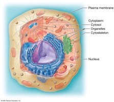
Most animal cells share three basic components: plasma membrane, cytoplasm, and nucleus. The cytoplasm includes cytosol, organelles, and cytoskeleton.
Plasma Membrane: Boundary separating the cell from its environment; regulates transport, communication, and identification.
Cytoplasm: Contains cytosol (intracellular fluid), organelles, and cytoskeleton.
Nucleus: Contains most of the cell's DNA and is the site for RNA production.
Cell Size and Diversity
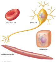
Cells vary greatly in size and appearance, enabling specialized functions throughout the human body.
Examples: Red blood cells, nerve cells, epithelial cells, and skeletal muscle cells.
The Plasma Membrane
The Phospholipid Bilayer
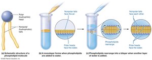
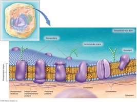
The plasma membrane is primarily composed of a phospholipid bilayer, forming a selective barrier between the extracellular fluid (ECF) and cytosol.
Hydrophilic (polar) heads: Face water.
Hydrophobic (nonpolar) tails: Repel water.
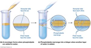
Phospholipids rearrange into two layers to exclude water from the fatty acid tails.
The Fluid Mosaic Model
The plasma membrane is a dynamic structure with proteins, lipids, and carbohydrates embedded within the bilayer, allowing fluidity and mosaic-like arrangement.
Integral proteins: Span the membrane; transmembrane if reaching both sides.
Peripheral proteins: Located on one side; may be anchored or float freely.
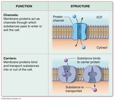
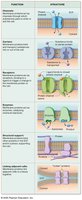
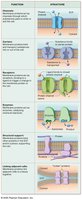
Membrane protein functions: Channels, carriers, receptors, enzymes, structural support, and linker proteins.
Other Membrane Components
Cholesterol: Stabilizes membrane structure during temperature changes.
Glycolipids and Glycoproteins: Carbohydrate chains attached to lipids/proteins; function in cell recognition.
Transport Across the Plasma Membrane
Passive Transport Processes
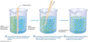
Passive transport does not require energy and relies on concentration gradients.
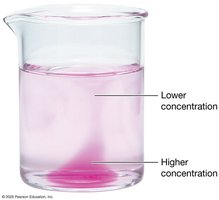
Diffusion: Movement of solute from high to low concentration.
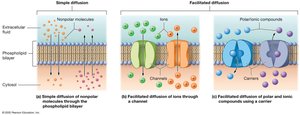
Simple Diffusion: Nonpolar solutes and gases pass directly through the bilayer.
Facilitated Diffusion: Polar/charged solutes cross via channel or carrier proteins.
Osmosis
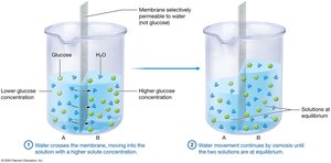
Osmosis is the movement of water across a selectively permeable membrane from lower to higher solute concentration.
Water crosses via aquaporins or between phospholipids.
Results in changes in fluid volume in compartments.
Tonicity
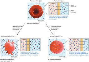
Tonicity compares solute concentrations and their ability to cause osmosis.
Isotonic: No net water movement.
Hypertonic: Cell loses water and shrivels (crenate).
Hypotonic: Cell gains water, swells, and may rupture (lyse).
Active Transport Processes
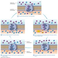
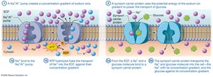
Active transport requires ATP to move solutes against their concentration gradient.
Primary Active Transport: Direct use of ATP; e.g., sodium-potassium pump moves 3 Na+ out and 2 K+ in.
Secondary Active Transport: Uses gradient created by primary transport to move another substance.
Vesicular Transport
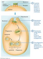
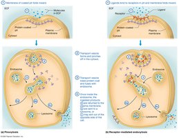
Large particles are transported via vesicles, requiring ATP.
Endocytosis: Bringing substances into the cell (phagocytosis, pinocytosis, receptor-mediated).
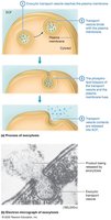
Exocytosis: Releasing substances from the cell.
Transcytosis: Substance moves through the cell and exits on the opposite side.
Summary Table: Plasma Membrane Transport
Comparison of transport mechanisms across the plasma membrane.
Transport Type | Energy Required | Direction | Example |
|---|---|---|---|
Simple Diffusion | No | High to Low | O2, CO2 |
Facilitated Diffusion | No | High to Low | Glucose, ions |
Osmosis | No | Water movement | Water |
Primary Active Transport | Yes (ATP) | Low to High | Na+/K+ pump |
Secondary Active Transport | Yes (ATP indirectly) | Coupled | Glucose absorption |
Vesicular Transport | Yes (ATP) | Bulk movement | Phagocytosis, exocytosis |
*Additional info: Table entries inferred for completeness based on textbook context.*
Cytoplasmic Organelles
Membrane-Enclosed Organelles
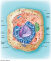
Organelles compartmentalize cellular functions, increasing efficiency and protecting cellular components.
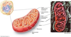
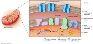
Mitochondria: ATP production; double membrane with cristae.
Peroxisomes: Oxidize toxic substances, break down fatty acids, synthesize phospholipids.
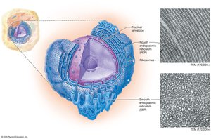
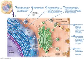
Endoplasmic Reticulum (ER): Rough ER (protein folding, export), Smooth ER (lipid synthesis, detoxification).
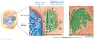
Golgi Apparatus: Modifies, sorts, and packages proteins/lipids.
Lysosomes: Digest macromolecules, autophagy, immune defense.
Non-Membrane-Enclosed Organelles
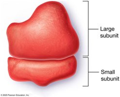
Ribosomes: Site of protein synthesis; free or bound.
Centrosome: Contains centrioles; organizes microtubules.
The Cytoskeleton
Types of Filaments
The cytoskeleton provides structural support, movement, and specialized functions.
Actin Filaments (Microfilaments): Structural support, tension, movement (muscle contraction).
Intermediate Filaments: Mechanical strength, support nuclear membrane, unite cells.
Microtubules: Internal architecture, organelle movement, form cilia and flagella.
The Nucleus
Structure and Function
The nucleus directs cellular activities and houses DNA, which contains genetic instructions for protein synthesis.
Nuclear Envelope: Double membrane with nuclear pores.
Nucleoplasm: Gel-like substance containing DNA and proteins.
Nucleolus: Site of ribosome assembly.
Chromatin and Chromosomes
DNA is organized as chromatin during interphase and condenses into chromosomes during cell division.
Chromatin: DNA and histone proteins; nucleosomes.
Chromosomes: Highly compacted DNA for cell division; humans have 46 chromosomes.
Protein Synthesis
Gene Expression
Protein synthesis involves transcription and translation, converting genetic information into functional proteins.
Transcription: DNA code copied to mRNA in the nucleus.
Translation: Ribosomes read mRNA and synthesize polypeptides.
Posttranslational Modification: Folding and alteration of polypeptides.
The Cell Cycle
Phases of the Cell Cycle
The cell cycle is the series of events from cell formation to division, essential for growth, development, and repair.
Interphase: Growth and preparation (G1, S, G2 phases).
S Phase: DNA replication via semiconservative mechanism.
M Phase: Mitosis (division of genetic material) and cytokinesis (division of cytoplasm).
Cell Cycle Control and Cancer
Cell division is regulated by checkpoints and signals; failure of control can lead to cancer.
Checkpoints: Monitor cell cycle progression.
Apoptosis: Programmed cell death for damaged or unnecessary cells.
Tumors: Benign (localized) or malignant (cancerous, can metastasize).
Summary
The cell is a complex, dynamic unit that performs essential functions for life. Understanding cell structure, membrane transport, organelles, protein synthesis, and the cell cycle is foundational for anatomy and physiology students.
