 뒤로
뒤로The Cellular Level of Organization: Structure and Function of Cells
스터디 가이드 - 스마트 노트
 자료에 맞춘 맞춤형 노트, 핵심 정의, 예시, 맥락을 확장해 제공합니다.
Chapter 3: The Cellular Level of Organization
Cell Theory and Introduction to Cells
Cell theory is a foundational concept in biology, stating that all living organisms are composed of cells, which arise from the division of preexisting cells. Cells are the smallest units capable of performing all vital physiological functions, and each cell maintains homeostasis at the cellular level.
Cytology: The study of cells, a branch of cell biology.
Somatic cells: All body cells except sex cells; undergo mitosis.
Sex cells (germ cells): Sperm in males and oocytes in females; undergo meiosis.
3-1 Plasma Membrane
Structure and Function of the Plasma Membrane
The plasma membrane, also known as the cell membrane, separates the cytoplasm from the extracellular fluid and is essential for maintaining cellular integrity and function.
Extracellular fluid (interstitial fluid): Watery medium surrounding the cell; about 1/3 of body fluid.
Intracellular fluid (cytoplasm): Fluid inside the cell; about 2/3 of body fluid.
Functions of the plasma membrane:
Physical isolation (barrier)
Regulation of exchange with the environment (ions, nutrients, wastes)
Sensitivity to the environment (chemical signals, extracellular fluid composition)
Structural support (anchors cells and tissues)
Membrane Lipids: The Phospholipid Bilayer
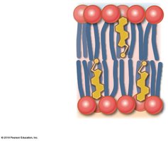
The plasma membrane is primarily composed of a phospholipid bilayer, which forms a barrier to ions and water-soluble compounds.
Hydrophilic heads: Face outward toward watery environments.
Hydrophobic fatty-acid tails: Face inward, away from water.
Membrane Proteins
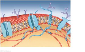
Membrane proteins are critical for the diverse functions of the plasma membrane.
Integral proteins: Embedded within the membrane.
Peripheral proteins: Bound to the inner or outer surface.
Types of membrane proteins:
Anchoring proteins: Attach to structures inside or outside the cell.
Recognition proteins: Label cells as normal or abnormal.
Enzymes: Catalyze reactions.
Receptor proteins: Bind and respond to ligands (e.g., hormones).
Carrier proteins: Transport specific solutes across the membrane.
Channels: Regulate water flow and solute passage; may be gated.
Membrane Carbohydrates: The Glycocalyx
Carbohydrates on the cell surface form the glycocalyx, a sticky "sugar coat" composed of proteoglycans, glycoproteins, and glycolipids.
Functions of the glycocalyx:
Lubrication and protection
Anchoring and locomotion
Specificity in binding (receptors)
Recognition (immune response)
3-2 Organelles within the Cytoplasm
Cytoplasm and Its Components
The cytoplasm includes all materials inside the cell, excluding the nucleus. It consists of cytosol (intracellular fluid), organelles, and inclusions.
Cytosol: Contains dissolved nutrients, ions, proteins, and waste products; high in protein and potassium.
Organelles: Structures with specific functions.
Inclusions: Masses of insoluble materials (e.g., glycogen, lipid droplets, melanin).
Types of Organelles
Nonmembranous organelles: Direct contact with cytosol (e.g., cytoskeleton, microvilli, centrioles, cilia, flagella, ribosomes, proteasomes).
Membranous organelles: Isolated from cytosol by a membrane (e.g., endoplasmic reticulum, Golgi apparatus, lysosomes, peroxisomes, mitochondria).
The Cytoskeleton
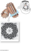
The cytoskeleton provides structural support, shape, and movement for the cell. It consists of microfilaments, intermediate filaments, and microtubules.
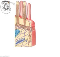
Microfilaments: Smallest, composed of actin; support cell shape.
Intermediate filaments: Provide strength and stabilize organelles.
Microtubules: Largest, form spindle apparatus during cell division.
Microvilli, Cilia, and Flagella
These cell surface structures are specialized for movement and increasing surface area.
Microvilli: Increase surface area for absorption (e.g., in the small intestine); attached to cytoskeleton via microfilaments.
Cilia: Motile extensions that move fluids across the cell surface; found in respiratory and reproductive tracts.
Flagella: Whip-like extension for cell movement; present only in sperm cells.
Ribosomes and Proteasomes
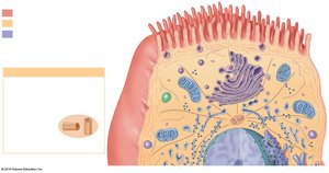
Ribosomes: Organelles that synthesize proteins; composed of rRNA and proteins. Free ribosomes produce proteins for the cytosol, while fixed ribosomes (on rough ER) produce proteins for secretion or membrane insertion.
Proteasomes: Contain proteases for breaking down damaged or abnormal proteins.
Membranous Organelles
Endoplasmic Reticulum (ER): Network of membranes with cisternae; involved in synthesis, storage, transport, and detoxification.
Smooth ER (SER): Synthesizes lipids and carbohydrates.
Rough ER (RER): Surface covered with ribosomes; synthesizes proteins and glycoproteins.
Golgi Apparatus: Modifies, packages, and sorts proteins and lipids for secretion or delivery to other organelles.
Lysosomes: Vesicles containing digestive enzymes; break down waste, bacteria, and damaged organelles.
Peroxisomes: Vesicles with enzymes that break down fatty acids and neutralize hydrogen peroxide.
Mitochondria: Produce ATP through aerobic metabolism; have a double membrane with inner folds (cristae).
3-3 Cell Nucleus
Structure and Function of the Nucleus
The nucleus is the largest organelle and serves as the cell's control center. It is surrounded by a double membrane (nuclear envelope) with nuclear pores for communication.
Nucleolus: Site of rRNA synthesis and ribosome assembly.
Chromatin: Loosely coiled DNA in non-dividing cells; condenses into chromosomes during cell division.
Genetic code: DNA sequence that determines protein structure; organized into genes (functional units of heredity).
3-4 Protein Synthesis
Gene Expression: Transcription and Translation
Protein synthesis is the process by which cells build proteins based on genetic instructions.
Transcription: DNA is used as a template to synthesize messenger RNA (mRNA).
RNA processing: Introns are removed, and exons are spliced together before mRNA exits the nucleus.
Translation: mRNA binds to ribosomes in the cytoplasm, where transfer RNA (tRNA) brings amino acids to build the polypeptide chain according to the mRNA codons.
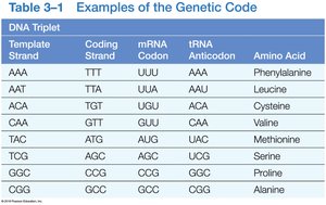
DNA Triplet | Coding Strand | mRNA Codon | tRNA Anticodon | Amino Acid |
|---|---|---|---|---|
AAA | TTT | UUU | AAA | Phenylalanine |
AAT | TTA | UUA | AAU | Leucine |
ACA | TGT | UGU | ACA | Cysteine |
CAA | GTT | GUU | CAA | Valine |
TAC | ATG | AUG | UAC | Methionine |
TCG | AGC | AGC | UCG | Serine |
GCG | CGC | CGC | GCG | Proline |
CGG | GCC | GCC | CGG | Alanine |
3-5 Diffusion and Osmosis
Membrane Transport Mechanisms
The plasma membrane is selectively permeable, allowing some substances to pass while restricting others. Transport can be passive (no energy required) or active (requires energy).
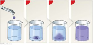
Diffusion: Movement of molecules from high to low concentration (down a concentration gradient).
Osmosis: Diffusion of water across a selectively permeable membrane toward higher solute concentration.
Factors Affecting Diffusion
Distance, molecular size, temperature, concentration gradient, and electrical forces.
Types of Diffusion Across Plasma Membranes
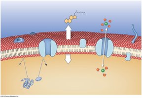
Simple diffusion: Direct passage of lipid-soluble molecules and gases through the membrane.
Channel-mediated diffusion: Passage of water-soluble compounds and ions through protein channels.
Osmolarity and Tonicity
Osmolarity: Total solute concentration in a solution (mOsM).
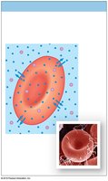
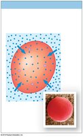
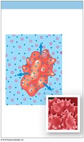
Tonicity: Effect of a solution on cell volume (isotonic, hypotonic, hypertonic).
Isotonic: No net water movement; cell remains unchanged.
Hypotonic: Water enters cell; cell may swell and burst (hemolysis).
Hypertonic: Water leaves cell; cell shrinks (crenation).
3-6 Carrier-Mediated and Vesicular Transport
Carrier-Mediated Transport
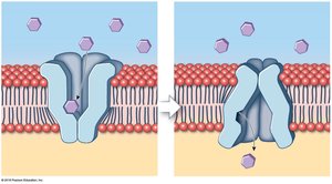
Facilitated diffusion: Passive transport of large molecules (e.g., glucose, amino acids) via carrier proteins.
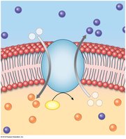
Active transport: Movement of substances against their concentration gradient using energy (ATP); includes ion pumps and exchange pumps (e.g., sodium-potassium pump).
Vesicular Transport
Endocytosis: Import of extracellular materials via vesicles (includes receptor-mediated endocytosis, pinocytosis, phagocytosis).
Exocytosis: Export of materials from the cell as vesicles fuse with the plasma membrane.
3-8 Cell Life Cycle
Cell Division
Mitosis: Produces two identical diploid daughter cells (46 chromosomes each).
Meiosis: Produces four unique haploid gametes (23 chromosomes each).
Interphase: Nondividing period; includes G0, G1, S, and G2 phases.
M phase: Mitosis (prophase, metaphase, anaphase, telophase) and cytokinesis (division of cytoplasm).
Cellular Differentiation
All cells contain the same genetic material but differentiate by turning off specific genes, leading to specialized cell functions (e.g., liver cells, neurons).
3-10 Cell Division and Cancer
Tumors and Cancer
Benign tumor: Contained, not life-threatening unless large.
Malignant tumor (cancer): Life-threatening due to metastasis (spread to other tissues).
Mutagens: Agents that cause mutations leading to cancer.
Summary Table: Types of Membrane Transport
Type | Energy Required? | Direction | Example |
|---|---|---|---|
Simple Diffusion | No | High to Low | O2, CO2 |
Facilitated Diffusion | No | High to Low | Glucose |
Active Transport | Yes (ATP) | Low to High | Na+/K+ pump |
Osmosis | No | Water: High to Low | Water movement |
Endocytosis/Exocytosis | Yes (ATP) | Bulk transport | Phagocytosis |
Additional info: This guide covers the essential concepts of cell structure, membrane transport, organelles, protein synthesis, and cell division, providing a foundation for further study in Anatomy & Physiology.
