 뒤로
뒤로The Cellular Level of Organization: Structure and Function of Cells
스터디 가이드 - 스마트 노트
 자료에 맞춘 맞춤형 노트, 핵심 정의, 예시, 맥락을 확장해 제공합니다.
The Cellular Level of Organization
Introduction to Cells
Cells are the fundamental structural and functional units of life. The study of cell structure is called cytology. According to cell theory, all living organisms are composed of cells, all cells arise from preexisting cells, and cells carry out essential physiological functions.
Somatic cells: All body cells except sex cells.
Sex cells (germ cells): Sperm in males and oocytes in females, responsible for reproduction.
Plasma Membrane
Structure and Functions
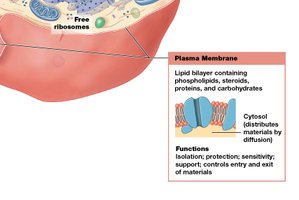
The plasma membrane forms the outer boundary of the cell and is essential for maintaining cellular integrity and function. It is primarily composed of a phospholipid bilayer with embedded proteins and carbohydrates.
Physical isolation: Separates the cell interior from the extracellular environment.
Regulation of exchange: Controls the entry and exit of ions, nutrients, and wastes.
Sensitivity: Contains receptors that detect chemical signals and environmental changes.
Structural support: Anchors cells to each other and stabilizes tissues.
Membrane Components
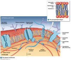
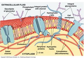
Lipids: The phospholipid bilayer has hydrophilic heads and hydrophobic tails. Cholesterol and glycolipids are also present, contributing to membrane fluidity and stability.
Proteins:
Integral proteins: Span the membrane and are involved in transport and signaling.
Peripheral proteins: Attached to the membrane surface, involved in signaling and maintaining cell shape.
Functional types: Anchoring, recognition, enzymes, receptors, carriers, and channels (including gated channels).
Carbohydrates: Glycoproteins, glycolipids, and proteoglycans form the glycocalyx, which provides lubrication, protection, and cell recognition.
Cellular Organelles
Types and Functions
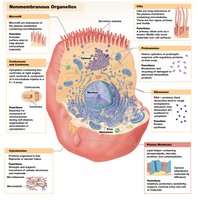
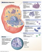
Organelles are specialized structures within the cytoplasm that perform distinct cellular functions. They are classified as nonmembranous or membranous.
Nonmembranous organelles: Cytoskeleton, centrioles, ribosomes, proteasomes, microvilli, cilia, and flagella.
Membranous organelles: Endoplasmic reticulum (ER), Golgi apparatus, lysosomes, peroxisomes, and mitochondria.
The Nucleus
Genetic Information and Function
The nucleus stores genetic information in the form of DNA. The genetic code consists of sequences of bases (A, T, C, G) that provide instructions for protein synthesis. A gene is a segment of DNA that codes for a specific protein.
Diffusion and Osmosis
Passive Transport Mechanisms
The plasma membrane is selectively permeable, allowing certain substances to pass while restricting others. Transport can be passive (no energy required) or active (requires energy).
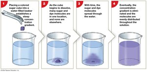
Diffusion: The net movement of molecules from an area of higher concentration to lower concentration, down a concentration gradient.
Osmosis: The diffusion of water across a selectively permeable membrane toward a higher solute concentration.
Factors Affecting Diffusion
Distance (shorter = faster)
Molecule size (smaller = faster)
Temperature (higher = faster)
Concentration gradient (steeper = faster)
Electrical forces (opposites attract, like charges repel)
Types of Diffusion Across Membranes
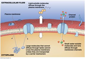
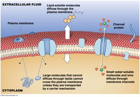
Simple diffusion: Lipid-soluble substances and gases cross the lipid bilayer directly.
Channel-mediated diffusion: Water and ions pass through protein channels.
Osmosis and Tonicity
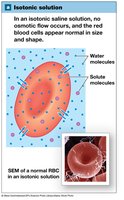
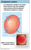
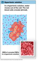
Osmotic pressure is the force with which water moves into a solution due to solute concentration. Hydrostatic pressure opposes osmotic pressure. Osmolarity is the total solute concentration in a solution. Tonicity describes how a solution affects cell volume:
Isotonic: No net water movement; cell remains unchanged.
Hypotonic: Water enters the cell; cell may swell and burst (hemolysis).
Hypertonic: Water leaves the cell; cell shrinks (crenation).
Carrier-Mediated and Vesicular Transport
Carrier-Mediated Transport
Carrier-mediated transport involves specialized membrane proteins and can be passive or active.
Specificity: Each carrier transports specific substances.
Saturation limits: Transport rate depends on carrier availability.
Regulation: Activity can be modified by cofactors such as hormones.
Symport (cotransport): Two substances move in the same direction.
Antiport (countertransport): Two substances move in opposite directions.
Facilitated Diffusion
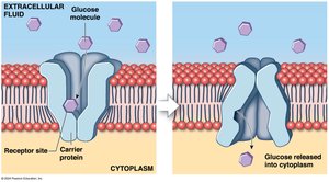
Facilitated diffusion is passive transport through carrier proteins. The transported molecule binds to a receptor site, causing the protein to change shape and allow passage.
Active Transport
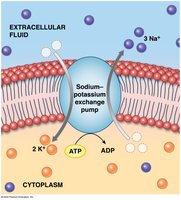
Active transport moves substances against their concentration gradients using energy (usually ATP). The sodium–potassium exchange pump is a primary example, moving three Na+ ions out and two K+ ions into the cell for each ATP consumed.
Primary active transport: Direct use of ATP (e.g., sodium–potassium pump).
Secondary active transport: Uses gradients established by primary active transport to move other substances.
Vesicular Transport
Vesicular (bulk) transport moves large particles or volumes via vesicles and requires ATP.
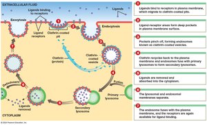
Endocytosis: Import of materials into the cell via vesicles.
Receptor-mediated endocytosis: Specific molecules are imported after binding to receptors.
Pinocytosis: Uptake of extracellular fluid.
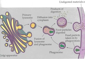
Phagocytosis: Uptake of solid particles.
Membrane Potential
Origin and Significance
The membrane potential is the electrical potential difference across the plasma membrane, resulting from the unequal distribution of positive and negative ions. The resting membrane potential of an unstimulated cell typically ranges from −10 mV to −100 mV. This potential is essential for nerve impulse transmission and muscle contraction.
The Cell Life Cycle
Stages of the Cell Cycle
The cell life cycle includes all events from one cell division to the next. It consists of interphase (G1, S, G2 phases) and the M phase (mitosis and cytokinesis).
Interphase: Cell grows, duplicates organelles (G1), replicates DNA (S), and synthesizes proteins (G2).
Mitosis: Division of the nucleus into two identical sets (prophase, metaphase, anaphase, telophase).
Cytokinesis: Division of the cytoplasm, producing two daughter cells.
Apoptosis: Programmed cell death.
Regulation of the Cell Life Cycle
Cell division is tightly regulated to balance cell loss and maintain tissue homeostasis. Regulation involves:
Stimulatory factors: Internal (e.g., M-phase promoting factor) and external (e.g., growth factors).
Inhibitory factors: Repressor genes and telomere shortening.
Cell Division and Cancer
Relationship Between Cell Division and Cancer
Cancer results from uncontrolled cell division due to mutations in genes that regulate cell growth and division. Tumors can be benign (localized) or malignant (invasive and metastatic). Mutated genes that drive cancer are called oncogenes, and agents that cause mutations are mutagens (including carcinogens such as chemicals, radiation, and certain pathogens).
Term | Definition |
|---|---|
Benign tumor | Remains in original tissue; rarely life-threatening |
Malignant tumor | Invades surrounding tissues; can metastasize |
Primary tumor | Original site of abnormal cell division |
Secondary tumor | Formed by metastasis from the primary tumor |
