 뒤로
뒤로The Cellular Level of Organization: Structure and Function of Cells
스터디 가이드 - 스마트 노트
 자료에 맞춘 맞춤형 노트, 핵심 정의, 예시, 맥락을 확장해 제공합니다.
The Cellular Level of Organization
Cell Theory and Types of Cells
The cell is the fundamental unit of life in all organisms. Cell theory states that all living things are composed of cells, cells arise from preexisting cells, and cells perform all vital physiological functions. Cells maintain homeostasis at the cellular level.
Cytology: The study of cells.
Sex cells (germ cells): Sperm (male) and oocytes (female).
Somatic cells: All other body cells except sex cells.
Anatomy of a Model Cell
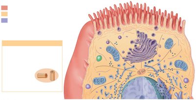
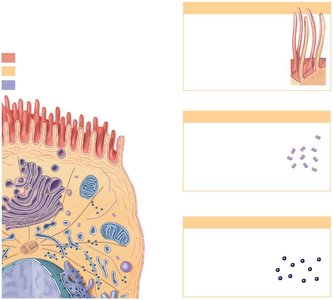
A typical cell contains various organelles, each with specific functions. Organelles are classified as either nonmembranous (direct contact with cytosol) or membranous (isolated from cytosol by a membrane).
Nonmembranous organelles: Cytoskeleton, centrioles, ribosomes, proteasomes, microvilli, cilia, flagella.
Membranous organelles: Endoplasmic reticulum (ER), Golgi apparatus, lysosomes, peroxisomes, mitochondria.
Cytoskeleton
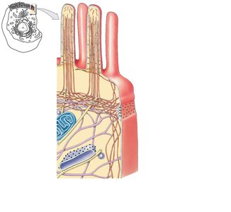
The cytoskeleton provides structural support, shape, and movement for the cell and its organelles. It consists of three main components:
Microfilaments: Thin filaments composed of actin; provide mechanical strength and interact with myosin for muscle contraction.
Intermediate filaments: Durable, mid-sized filaments; strengthen the cell and stabilize organelle position.
Microtubules: Large, hollow tubes of tubulin; anchor organelles, change cell shape, move organelles, form spindle apparatus, centrioles, and cilia.
Plasma Membrane Structure and Function
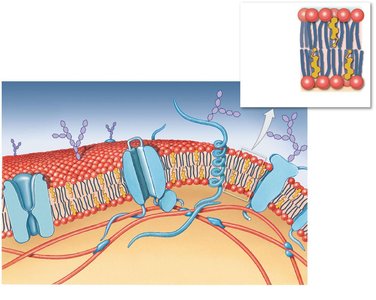
The plasma membrane separates the cytoplasm from the extracellular fluid and regulates the exchange of substances. It is composed of a phospholipid bilayer with embedded proteins and carbohydrates.
Phospholipid bilayer: Hydrophilic heads face outward; hydrophobic tails face inward, forming a barrier to ions and water-soluble compounds.
Membrane proteins: Integral (within membrane) and peripheral (bound to surface). Functions include anchoring, recognition, enzymatic activity, receptor binding, carrier transport, and channel formation.
Membrane carbohydrates: Proteoglycans, glycoproteins, glycolipids form the glycocalyx for lubrication, protection, anchoring, binding, and recognition.
Microvilli and Cilia
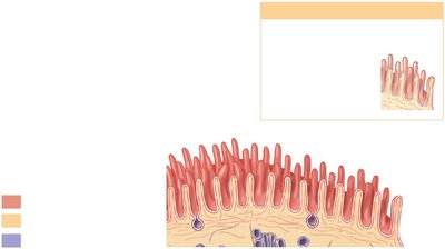
Microvilli and cilia are extensions of the plasma membrane that increase surface area or facilitate movement.
Microvilli: Increase surface area for absorption; contain microfilaments.
Cilia: Long extensions containing microtubules; primary cilia act as sensors, motile cilia move materials over cell surfaces.
Ribosomes and Proteasomes
Ribosomes are responsible for protein synthesis, while proteasomes break down damaged or abnormal proteins.
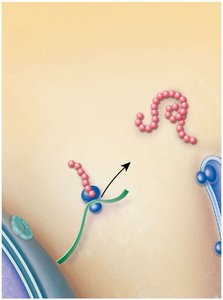
Ribosomes: Composed of RNA and proteins; free ribosomes synthesize proteins for cytosol, fixed ribosomes synthesize proteins for ER.
Proteasomes: Hollow cylinders with proteolytic enzymes; recycle damaged proteins.
Endoplasmic Reticulum (ER)
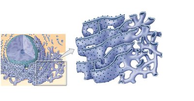
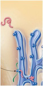
The ER is a network of membranous channels involved in synthesis, storage, transport, and detoxification.
Rough ER (RER): Covered with ribosomes; synthesizes and modifies proteins.
Smooth ER (SER): No ribosomes; synthesizes lipids, carbohydrates, steroid hormones, stores glycogen.
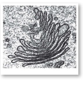
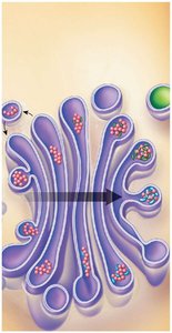
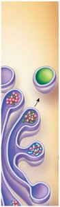
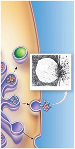
Golgi Apparatus
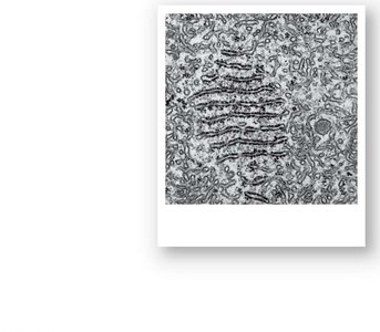
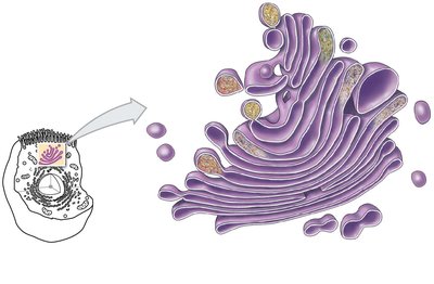
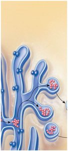
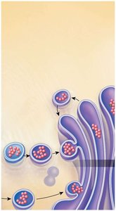
The Golgi apparatus modifies, packages, and sorts proteins and lipids for secretion or use within the cell.
Functions: Modifies secretions, adds/removes carbohydrates, renews plasma membrane, packages enzymes in lysosomes.
Lysosomes and Peroxisomes
Lysosomes and peroxisomes are vesicles containing enzymes for digestion and detoxification.
Lysosomes: Digest damaged organelles, pathogens; autolysis releases enzymes for cell destruction.
Peroxisomes: Break down fatty acids, neutralize toxic compounds; produce hydrogen peroxide, catalase converts it to water and oxygen.
Mitochondria
Mitochondria are the cell's powerhouses, producing ATP through aerobic metabolism.
Structure: Double membrane, inner folds (cristae) surround matrix.
Function: Glycolysis (cytosol), citric acid cycle (matrix), electron transport chain (inner membrane).
Aerobic metabolism:
Nucleus
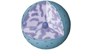
The nucleus is the largest organelle and the control center of the cell, containing genetic material.
Nuclear envelope: Double membrane with nuclear pores for communication.
Nucleoplasm: Contains nucleotides, enzymes, chromatin, nucleolus (site of rRNA synthesis).
Chromatin: Loosely coiled DNA; chromosomes form before cell division.
Genetic Code and Protein Synthesis
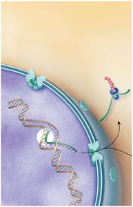
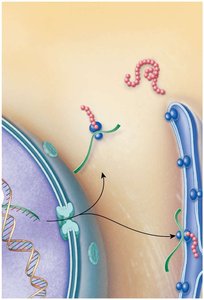
The genetic code is the sequence of DNA bases (A, T, C, G) that determines protein synthesis. Each gene codes for a specific protein.
Transcription: DNA is transcribed to mRNA in the nucleus.
Translation: mRNA is translated into a polypeptide at ribosomes in the cytoplasm.
Diffusion and Osmosis
Diffusion and osmosis are passive transport processes across the plasma membrane.
Diffusion: Movement from high to low concentration; affected by distance, size, temperature, gradient, and electrical forces.
Osmosis: Diffusion of water toward higher solute concentration; osmotic pressure is the force required to stop osmosis.
Tonicity: Effect of solution on cell size: isotonic (no change), hypotonic (cell swells), hypertonic (cell shrinks).
Carrier-Mediated and Vesicular Transport
Carrier-mediated transport uses proteins to move substances across the membrane. Vesicular transport moves materials in bulk.
Facilitated diffusion: Passive; carrier proteins transport large molecules (e.g., glucose).
Active transport: Requires ATP; moves substances against concentration gradient (e.g., sodium-potassium pump).
Vesicular transport: Endocytosis (receptor-mediated, pinocytosis, phagocytosis) and exocytosis.
Membrane Potential
Membrane potential is the electrical potential difference across the plasma membrane due to separation of charges. Resting membrane potential ranges from –10 mV to –100 mV depending on cell type.
Cell Life Cycle
The cell life cycle includes interphase (G0, G1, S, G2), mitosis, and cytokinesis. DNA replication occurs in S phase, mitosis divides the nucleus, and cytokinesis divides the cytoplasm.
Interphase: Cell growth, DNA replication, organelle duplication.
Mitosis: Prophase, metaphase, anaphase, telophase.
Cytokinesis: Division of cytoplasm into two daughter cells.
Regulation of Cell Cycle and Cancer
Cell division is regulated by internal and external factors. Cancer results from abnormal cell proliferation due to mutations in genes controlling cell growth.
Benign tumor: Contained, not life-threatening unless large.
Malignant tumor: Invades surrounding tissues, can metastasize.
Cellular Differentiation
All cells contain the same genetic material, but differentiate by turning off genes not needed for their specific function. This allows for specialized cell types such as liver cells, fat cells, and neurons.
Additional info: These notes provide a comprehensive overview of Chapter 3: The Cellular Level of Organization, covering cell structure, organelles, membrane transport, genetic code, protein synthesis, cell cycle, and differentiation, suitable for college-level Anatomy & Physiology students.
