뒤로
뒤로The Digestive System: Structure and Function
스터디 가이드 - 스마트 노트
 자료에 맞춘 맞춤형 노트, 핵심 정의, 예시, 맥락을 확장해 제공합니다.
The Digestive System: Overview
Main Functions of the Digestive System
The digestive system is responsible for the breakdown and absorption of nutrients, as well as the elimination of indigestible substances. Its main functions include:
Ingestion: Taking in food through the mouth.
Digestion: Breaking down food into nutrient molecules, both mechanically and chemically.
Absorption: Moving nutrient molecules into the bloodstream or lymphatic system.
Defecation: Eliminating indigestible remains as feces.
Organization of the Digestive System
Alimentary Canal and Accessory Organs
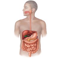
The digestive system is divided into the alimentary canal (gastrointestinal tract) and accessory digestive organs:
Alimentary Canal: A continuous muscular tube from mouth to anus, including the mouth, pharynx, esophagus, stomach, small intestine, large intestine, and anus.
Accessory Organs: Teeth, tongue, gallbladder, and digestive glands (salivary glands, liver, pancreas) that aid in digestion.
Digestive Processes
Six Essential Activities
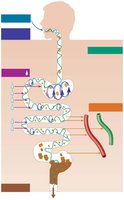
Food processing in the digestive system involves six essential activities:
Ingestion: Eating food.
Propulsion (Motility): Movement of food through the alimentary canal, including swallowing and peristalsis.
Mechanical Breakdown: Chewing, mixing, churning, and segmentation to physically break down food.
Digestion: Enzymatic breakdown of complex molecules into simpler chemical building blocks.
Absorption: Passage of digested fragments into blood or lymph.
Defecation: Elimination of indigestible substances as feces.
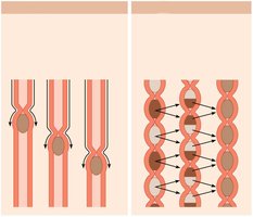
Peristalsis: Alternating waves of contraction and relaxation that propel food forward. Segmentation: Nonadjacent segments contract and relax, mixing food and breaking it down mechanically.
Histology of the Alimentary Canal
Four Basic Layers (Tunics)
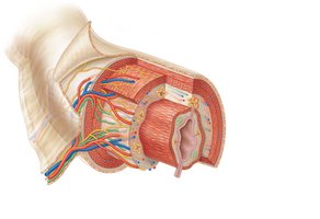
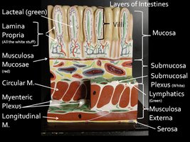
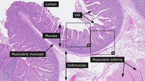
All digestive organs share a common structure with four basic layers:
Mucosa: Innermost layer lining the lumen; functions in secretion, absorption, and protection. Composed of epithelium, lamina propria, and muscularis mucosae.
Submucosa: Connective tissue containing blood vessels, lymphatics, and nerves.
Muscularis Externa: Responsible for segmentation and peristalsis; consists of inner circular and outer longitudinal muscle layers.
Serosa: Outermost layer; visceral peritoneum. In the esophagus, replaced by fibrous adventitia.
Blood Supply and Innervation
Splanchnic Circulation and Enteric Nervous System
Splanchnic Circulation: Arteries branching from the aorta supply digestive organs; hepatic portal circulation delivers nutrient-rich blood to the liver for processing.
Enteric Nervous System: The GI tract's own nervous system ("gut brain") regulates motility and secretion via the myenteric and submucosal nerve plexuses. It can function independently but is modulated by the central nervous system.
The Mouth and Associated Structures
Anatomy of the Oral Cavity
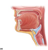
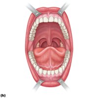
The mouth (oral cavity) is the entry point for food and is lined with stratified squamous epithelium for protection. The palate forms the roof of the mouth and is divided into the hard palate (bony) and soft palate (muscular, ending in the uvula).
The Tongue
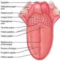
The tongue is a muscular organ that manipulates food, forms the bolus, and initiates swallowing, speech, and taste. It is anchored to the floor of the mouth by the lingual frenulum.
Salivary Glands
Major Salivary Glands and Functions of Saliva
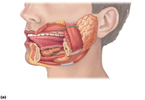
Saliva cleanses the mouth, dissolves food chemicals for taste, moistens food, and begins starch digestion with amylase. The three major salivary glands are:
Parotid Gland: Anterior to the ear, opens near the second upper molar.
Submandibular Gland: Medial to the mandible.
Sublingual Gland: Under the tongue, anterior in the mouth floor.
The Teeth
Types and Functions of Teeth
Teeth are essential for mastication (chewing), which is a form of mechanical digestion. Humans have two sets of teeth:
Deciduous (Primary) Teeth: 20 teeth that erupt between 6 and 24 months of age.
Permanant (Adult) Teeth: 32 teeth, including incisors (cutting), canines (tearing), premolars (grinding/crushing), and molars (grinding).
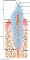
Tooth Structure
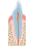
Each tooth consists of:
Crown: Exposed part above the gum, covered by enamel (hardest substance in the body).
Root: Embedded in the jawbone, covered by cementum, and attached to the periodontal ligament.
Dentin: Bonelike material under enamel, forms tubules.
Pulp Cavity: Contains connective tissue, blood vessels, and nerves.
Root Canal: Extension of the pulp cavity into the root.
Tooth and Gum Disease
Dental Caries, Gingivitis, and Periodontitis
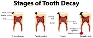
Dental Caries (Cavities): Demineralization of enamel and dentin by bacterial acids; prevented by good oral hygiene.
Gingivitis: Inflammation of the gums due to plaque and calculus; reversible if treated early.
Periodontitis: Advanced gum disease where immune cells attack tissues, potentially leading to tooth loss and increased risk of heart disease and stroke.