 뒤로
뒤로The Endocrine System: Structure, Function, and Clinical Correlations
스터디 가이드 - 스마트 노트
 자료에 맞춘 맞춤형 노트, 핵심 정의, 예시, 맥락을 확장해 제공합니다.
Endocrine System Overview
Introduction to the Endocrine System
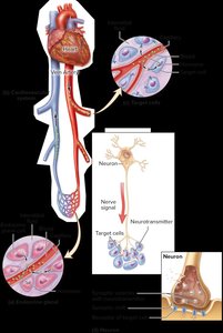
The endocrine system is a network of ductless glands that synthesize and secrete hormones, which are chemical messengers released into the blood and transported throughout the body. These hormones regulate a wide range of physiological processes by binding to specific receptors on target cells, enabling the cells to respond appropriately.
Hormones: Chemical messengers released into the bloodstream.
Target cells: Cells with specific receptors for a hormone, allowing them to respond to hormonal signals.
Transport: Hormones are released into interstitial fluid, enter the blood, and bind to receptors on target cells after leaving the bloodstream.
Hormone Effects
Hormones can change the types, quantities, or activities of enzymes and structural proteins in target cells. They can alter metabolic activities of multiple tissues and organs simultaneously and affect long-term processes such as growth and development.
Comparison of Endocrine and Nervous Systems
Control Systems of the Body
The endocrine and nervous systems are the two main control systems of the body. Both release ligands (chemical messengers) that bind to cellular receptors on target cells. However, there are key differences:
Endocrine system: Transmits hormones through the blood, targets any cell with the correct receptor, has widespread effects, longer reaction times, and longer-lasting effects (minutes to weeks).
Nervous system: Uses neurotransmitters, has localized and rapid effects, and shorter duration of action.
Both systems are mainly regulated by negative feedback mechanisms.
General Functions of the Endocrine System
Regulating development, growth, and metabolism: Hormones regulate embryonic cell division and metabolism.
Maintaining blood composition and volume: Hormones regulate blood solute concentrations, volume, cellular concentration, and platelet number.
Controlling digestive processes: Hormones influence secretory processes and movement of materials in the digestive tract.
Controlling reproductive activities: Hormones affect development and function of reproductive systems and sexual behaviors.
Major Endocrine Glands and Organs
Location and Structure
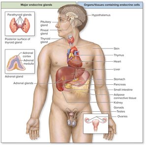
Endocrine glands contain epithelial tissue that produces and releases hormones within a connective tissue framework. Some glands are solely endocrine in function, while others contain clusters of endocrine cells within organs that have additional functions.
Primary endocrine organs: Pituitary, pineal, thyroid, parathyroid, and adrenal glands.
Other organs with endocrine cells: Hypothalamus, skin, thymus, heart, liver, stomach, pancreas, small intestine, adipose tissue, kidneys, and gonads.
Regulation of Hormone Synthesis and Release
Endocrine Reflexes
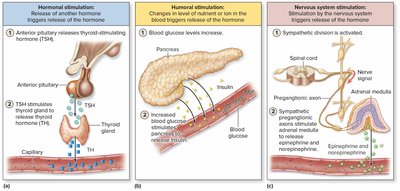
Hormone release is regulated by endocrine reflexes, which can be initiated by three types of stimuli:
Hormonal stimulation: Release of a hormone in response to another hormone.
Humoral stimulation: Changes in the level of a nutrient or ion in the blood trigger hormone release.
Nervous system stimulation: Stimulation by the nervous system triggers hormone release.
Classification of Hormones
Chemical Classes of Circulating Hormones
Hormones are classified based on their chemical structure:
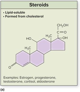
Steroids: Lipid-soluble molecules synthesized from cholesterol (e.g., estrogen, progesterone, testosterone, cortisol, aldosterone).
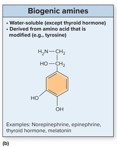
Biogenic amines (monoamines): Modified amino acids, mostly water-soluble except for thyroid hormone (e.g., norepinephrine, epinephrine, thyroid hormone, melatonin).
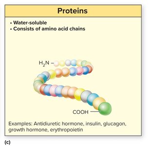
Proteins: Water-soluble chains of amino acids (e.g., antidiuretic hormone, insulin, glucagon, growth hormone, erythropoietin).
Local Hormones
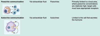
Local hormones are signaling molecules that do not circulate in the blood. They act on the cells that release them (autocrine stimulation) or on neighboring cells (paracrine stimulation). Eicosanoids, such as prostaglandins, are a type of local hormone formed from fatty acids and are involved in pain and inflammatory responses.
Hormone Transport in the Blood
Lipid-Soluble vs. Water-Soluble Hormones
Lipid-soluble hormones: Require carrier proteins for transport in the blood. Only unbound (free) hormone can exit the blood and bind to target cell receptors.
Water-soluble hormones: Most travel freely in the blood; a few use carrier proteins to prolong their half-life.
Hormone Action on Target Cells
Mechanisms of Hormone Action
Lipid-soluble hormones: Diffuse across the target cell membrane and bind to intracellular receptors.
Water-soluble hormones: Bind to cell-surface receptors, initiating a signal transduction pathway involving second messengers (e.g., cAMP, Ca2+).
Amplification: One hormone-receptor interaction can activate many second messengers, magnifying the effect.
Receptor cascade: Activation of multiple second messengers leads to diverse cellular effects.
Regulation of Target Cell Sensitivity
Receptor Number and Regulation
Down-regulation: Decrease in the number of hormone receptors in response to high hormone levels, reducing cell sensitivity.
Up-regulation: Increase in the number of hormone receptors in response to low hormone levels, increasing cell sensitivity.
A cell’s response depends on receptor number and simultaneous exposure to other hormones.
Hormone Interactions
Synergistic: One hormone reinforces the activity of another (e.g., estrogen and progesterone).
Permissive: One hormone requires the activity of another (e.g., oxytocin’s milk ejection requires prolactin’s milk production).
Antagonistic: One hormone opposes the activity of another (e.g., glucagon increases blood glucose, insulin lowers it).
Integrative: Hormones produce different but complementary results.
The Hypothalamus and Pituitary Gland
Anatomic Relationship
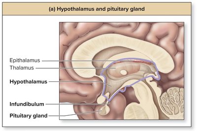
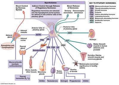
The hypothalamus controls the pituitary gland, which in turn regulates several other endocrine organs. The pituitary gland is located inferior to the hypothalamus and is connected by the infundibulum. It is divided into anterior and posterior lobes.
Posterior Pituitary
The posterior pituitary stores and releases antidiuretic hormone (ADH) and oxytocin (OT), both produced by the hypothalamus.
ADH (vasopressin): Decreases urine production, stimulates thirst, constricts blood vessels.
Oxytocin: Stimulates uterine contraction, milk ejection, and emotional bonding.
Anterior Pituitary
The anterior pituitary is hormonally stimulated by the hypothalamus to release its own hormones into general circulation.
Releasing hormones (RHs): Increase secretion of anterior pituitary hormones (e.g., TRH, PRH, GnRH, CRH, GHRH).
Inhibiting hormones (IHs): Decrease secretion of anterior pituitary hormones (e.g., PIH, GIH).
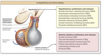
Anterior Pituitary Hormones
Thyroid-stimulating hormone (TSH): Stimulates thyroid hormone release.
Prolactin (PRL): Stimulates milk production and mammary gland growth.
Adrenocorticotropic hormone (ACTH): Stimulates corticosteroid release from the adrenal cortex.
Gonadotropins (FSH and LH): Regulate reproductive functions in both sexes.
Growth hormone (GH): Stimulates cell growth, division, and the release of insulin-like growth factors (IGFs).
Growth Hormone: Regulation and Effects
Regulation of GH Release
Stimulated by age (highest in children/adolescents), time of day (peaks at night), nutrient levels, stress, and exercise.
Severe or chronic stress can decrease GH.
Effects of GH and IGFs
Stimulate protein synthesis, cell division, and differentiation.
Increase glycogenolysis and gluconeogenesis; inhibit glycogenesis.
Stimulate lipolysis; inhibit lipogenesis.
Disorders of GH Secretion
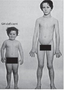
Growth hormone deficiency (pituitary dwarfism): Short stature, low blood sugar.
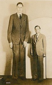
Pituitary gigantism: Excessive growth, increased blood sugar, enlarged organs.
Acromegaly: Excess GH in adults, enlargement of bones in face, hands, feet, and increased glucose release.
The Thyroid Gland
Anatomy and Histology
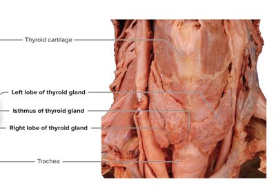
The thyroid gland is located inferior to the thyroid cartilage of the larynx and anterior to the trachea. It consists of left and right lobes connected by an isthmus and is highly vascularized.
Follicular cells: Synthesize thyroglobulin and produce thyroid hormone (TH).
Parafollicular cells: Produce calcitonin, which decreases blood calcium levels.
Colloid: Viscous, protein-rich fluid in the follicle lumen.
Thyroid Hormone Regulation and Effects
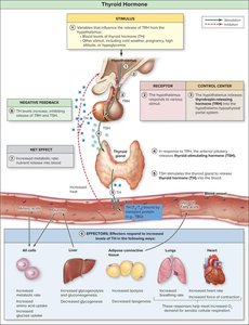
Hypothalamus releases TRH → anterior pituitary releases TSH → thyroid releases TH (T3 and T4).
TH increases metabolic rate and nutrient release into blood.
Regulated by negative feedback; set point influenced by genetics, age, and environment.
Disorders of Thyroid Hormone Secretion
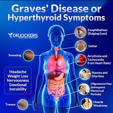
Hyperthyroidism: Excess TH, increased metabolic rate, weight loss, hyperactivity, heat intolerance. Causes include Graves disease, excessive T4, or pituitary overstimulation.
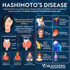
Hypothyroidism: Decreased TH, low metabolic rate, lethargy, cold intolerance, weight gain. Causes include iodine deficiency, pituitary dysfunction, or autoimmune destruction (Hashimoto thyroiditis).
Goiter: Thyroid enlargement, usually due to iodine deficiency.

Calcitonin: Regulation and Effects
Produced by parafollicular cells in response to high blood calcium or exercise.
Decreases blood calcium by inhibiting osteoclasts and increasing calcium excretion in urine.
Most significant in children due to high bone turnover.
The Adrenal Glands
Anatomy and Regions
The adrenal glands are located on the superior surface of each kidney and consist of two regions: the adrenal medulla (inner core) and the adrenal cortex (outer layer).
Adrenal medulla: Releases epinephrine and norepinephrine during sympathetic stimulation.
Adrenal cortex: Produces over 25 corticosteroids in three zones:
Zona glomerulosa: Mineralocorticoids (e.g., aldosterone) regulate electrolytes.
Zona fasciculata: Glucocorticoids (e.g., cortisol) regulate blood sugar.
Zona reticularis: Gonadocorticoids (sex hormones).
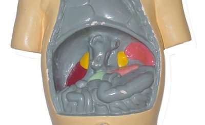
Cortisol: Regulation and Effects
Regulated by negative feedback, time of day (peaks in early morning), and stress.
Increases blood nutrient levels by stimulating glycogenolysis, gluconeogenesis, and lipolysis; inhibits glycogenesis and lipogenesis.
High doses suppress inflammation and immune response but can cause side effects (e.g., infection risk, sodium retention).
Disorders of Adrenal Cortex Hormone Secretion
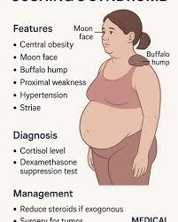
Cushing syndrome: Excess glucocorticoids, causing obesity, hypertension, hirsutism, kidney stones, and menstrual irregularities.
Addison disease: Adrenal insufficiency, causing weight loss, fatigue, hypotension, and skin darkening.
Adrenogenital syndrome: Excess androgen production, leading to masculinization in newborns.
Stress Response (General Adaptation Syndrome)
Alarm reaction: Immediate sympathetic activation (fight or flight), epinephrine, norepinephrine release.
Stage of resistance: Cortisol secretion maintains blood sugar and energy supply.
Stage of exhaustion: Prolonged stress depletes energy stores, leading to protein breakdown and illness.
The Pancreas
Anatomy and Function
The pancreas is located posterior to the stomach and has both endocrine and exocrine functions. Acinar cells produce digestive enzymes, while pancreatic islets contain endocrine cells:
Alpha cells: Secrete glucagon (raises blood glucose).
Beta cells: Secrete insulin (lowers blood glucose).
Delta cells: Secrete somatostatin.
F cells: Secrete pancreatic polypeptide.
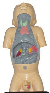
Clinical Correlations: Glucose Regulation Disorders
Diabetes mellitus: Inadequate glucose uptake, leading to chronic hyperglycemia and complications (blindness, kidney failure, amputations).
Type 1 diabetes: Autoimmune destruction of beta cells; requires insulin therapy.
Type 2 diabetes: Insulin resistance, often associated with obesity; managed with diet, exercise, and medication.
Gestational diabetes: Occurs during pregnancy; increases risk of type 2 diabetes later.
Hypoglycemia: Low blood glucose, caused by insulin overdose, prolonged exercise, or organ dysfunction; treated with glucagon if unconscious.
Pineal and Parathyroid Glands
Pineal Gland
Located in the epithalamus; secretes melatonin at night.
Regulates circadian rhythm and mood; influences GnRH secretion.
Parathyroid Glands
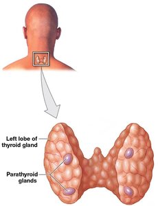
Small glands on the posterior thyroid; usually four in number.
Chief cells: Produce parathyroid hormone (PTH), which increases blood calcium by stimulating bone resorption, decreasing urinary loss, and activating calcitriol.
Oxyphil cells: Function unknown.
Other Endocrine Structures
Thymus: Secretes thymic hormones for T-lymphocyte maturation; shrinks with age.
Heart: Atrial natriuretic peptide (ANP) lowers blood pressure by increasing urine output and dilating vessels.
Kidneys: Release erythropoietin (EPO) to stimulate red blood cell production.
Liver: Secretes insulin-like growth factors and angiotensinogen (precursor to angiotensin II, which raises blood pressure).
Stomach: Secretes gastrin to increase digestive activity.
Small intestine: Secretes secretin and cholecystokinin (CCK) to regulate digestion.
Skin: Produces vitamin D3, converted to calcitriol to raise blood calcium.
Adipose tissue: Secretes leptin to regulate appetite; excess or deficiency affects puberty and menstrual cycles.
Aging and the Endocrine System
Secretory activity of endocrine glands decreases with age, reducing hormone levels and efficiency of endocrine functions.
Decreased growth hormone and sex hormones contribute to loss of weight and body mass in the elderly.
