 뒤로
뒤로The Endocrine System: Structure, Function, and Regulation
스터디 가이드 - 스마트 노트
 자료에 맞춘 맞춤형 노트, 핵심 정의, 예시, 맥락을 확장해 제공합니다.
Chapter 16: The Endocrine System
Overview of the Endocrine System
The endocrine system is one of the two major regulatory systems of the body, alongside the nervous system. It consists of glands that synthesize and secrete chemical messengers called hormones into the bloodstream. These hormones interact with specific target cells that possess receptors for the hormone, leading to changes in cellular function. The tissues containing these target cells are known as target tissues.
Hormones: Chemical messengers secreted into the blood to regulate physiological pro ce s ses.
Target Cells: Cells with specific receptors for a hormone.a I
Receptors: Proteins on or in target cells that bind hormones and initiate cellular changes.
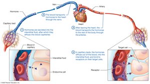
Comparison of the Endocrine and Nervous Systems
The endocrine and nervous systems both regulate body functions but differ in their mechanisms and effects:
Endocrine system: Hormones are secreted into the interstitial fluid, diffuse into blood capillaries, and are transported throughout the body. Effects are generally slower to initiate but longer-lasting.
Nervous system: Neurotransmitters are released directly onto target cells, producing rapid but short-lived effects.
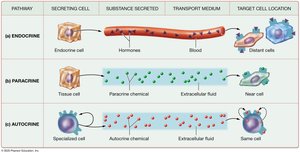
Types of Chemical Signaling
Not all chemical signals are classic hormones. Other types include:
Paracrine signals: Chemicals secreted into the extracellular fluid to affect nearby cells.
Autocrine signals: Chemicals secreted by a cell that affect the same cell.
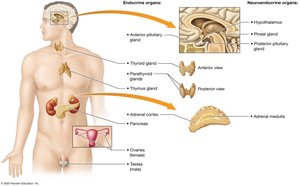
Endocrine Organs
Endocrine glands are ductless organs that secrete hormones into the interstitial fluid for transport by the bloodstream. In contrast, exocrine glands secrete their products into ducts leading to body surfaces or cavities.
Primary endocrine organs: Anterior pituitary, thyroid, parathyroid, adrenal cortices, pancreas, thymus, ovaries/testes.
Secondary endocrine glands: Heart, kidneys, small intestine, adipose tissue.
Neuroendocrine organs: Hypothalamus, pineal gland, adrenal medulla (nervous tissue that secretes hormones).
Hormones: Structure, Function, and Mechanisms
Classes of Hormones
Amino acid-based hormones: Derived from amino acids; generally hydrophilic (except thyroid hormone, which is hydrophobic).
Peptide/protein hormones: Chains of amino acids; hydrophilic.
Steroid hormones: Derived from cholesterol; hydrophobic and lipid-soluble.
Hormone Transport in Blood
Free hormones: Hydrophilic, travel unbound in plasma.
Bound hormones: Hydrophobic, travel bound to plasma proteins, which extends their half-life.
Target Cells and Receptors
Hormones bind to specific receptors on or in target cells. The number of receptors can be regulated:
Upregulation: Increase in receptor number in response to low hormone levels.
Downregulation: Decrease in receptor number after prolonged exposure to high hormone levels.
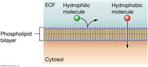
Mechanisms of Hormone Action
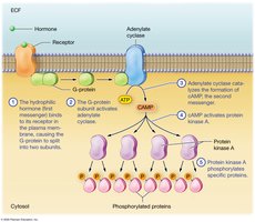
Hydrophilic hormones: Bind to cell surface receptors and activate second-messenger systems (e.g., cAMP pathway).
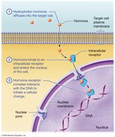
Hydrophobic hormones: Diffuse through the plasma membrane, bind to intracellular receptors, and directly influence gene expression.
Regulation of Hormone Secretion
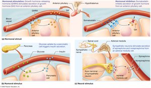
Hormone secretion is regulated by three main types of stimuli:
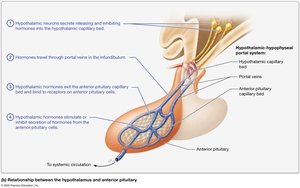
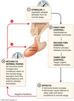
Hormonal stimuli: Hormones stimulate the release of other hormones (e.g., hypothalamic hormones regulate anterior pituitary hormones).
Humoral stimuli: Changes in blood levels of ions or nutrients trigger hormone release (e.g., blood glucose regulates insulin).
Neural stimuli: Nerve fibers stimulate hormone release (e.g., sympathetic stimulation of adrenal medulla).
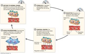
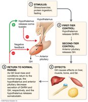
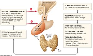
Most hormone secretion is regulated by negative feedback loops, maintaining homeostasis.
Hypothalamus and Pituitary Gland
Structure and Functional Relationships
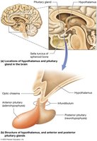
The hypothalamus connects to the pituitary gland via the infundibulum. The pituitary gland has two parts:
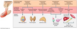
Anterior pituitary (adenohypophysis): True glandular tissue; secretes hormones in response to hypothalamic releasing/inhibiting hormones via the hypophyseal portal system.
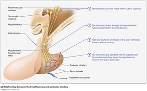
Posterior pituitary (neurohypophysis): Nervous tissue; stores and releases hormones produced by the hypothalamus (ADH and oxytocin).
Hormones of the Posterior Pituitary
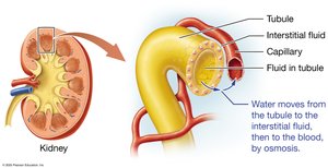
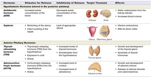
Antidiuretic hormone (ADH): Promotes water retention by the kidneys; released in response to high blood solute concentration.
Oxytocin: Stimulates uterine contractions and milk ejection; involved in positive feedback during childbirth and lactation.
Hormones of the Anterior Pituitary
Thyroid-stimulating hormone (TSH): Stimulates thyroid hormone production.
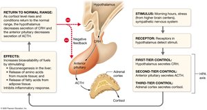
Adrenocorticotropic hormone (ACTH): Stimulates adrenal cortex hormone production.
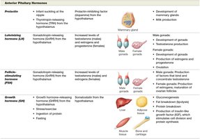
Prolactin: Stimulates milk production.
Luteinizing hormone (LH) and Follicle-stimulating hormone (FSH): Regulate gonadal function.
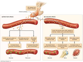
Growth hormone (GH): Stimulates growth, protein synthesis, and metabolism.
Summary Table: Hypothalamic and Pituitary Hormones
Thyroid and Parathyroid Glands
Structure and Function
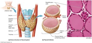
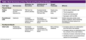
Thyroid gland: Located in the anterior neck; produces thyroid hormones (T3 and T4) and calcitonin.
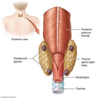
Parathyroid glands: Usually four glands on the posterior thyroid; produce parathyroid hormone (PTH).
Thyroid Hormones
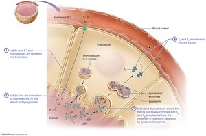
Triiodothyronine (T3) and Thyroxine (T4): Regulate basal metabolic rate, thermoregulation, growth, and development; synergize with the sympathetic nervous system.
Production involves iodination of thyroglobulin in the colloid of thyroid follicles.
Thyroid Disorders
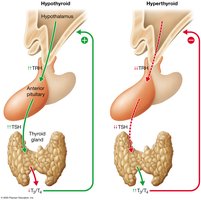
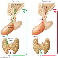
Hyperthyroidism: Excess thyroid hormone; causes weight loss, heat intolerance, and increased heart rate.
Hypothyroidism: Deficient thyroid hormone; causes weight gain, cold intolerance, and slow heart rate.
Graves disease: Autoimmune hyperthyroidism; may cause goiter and exophthalmos.
Goiter: Enlargement of the thyroid gland.

Parathyroid Hormone and Calcitonin
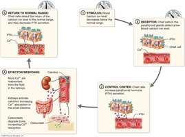
Parathyroid hormone (PTH): Increases blood calcium by stimulating osteoclasts, increasing intestinal absorption, and increasing renal reabsorption of calcium.
Calcitonin: Lowers blood calcium by inhibiting osteoclasts; secreted by thyroid parafollicular cells in response to hypercalcemia.
Adrenal Glands
Structure
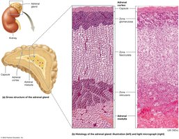
The adrenal glands are located on the superior aspect of each kidney and consist of an outer cortex and inner medulla.
Adrenal cortex: Produces steroid hormones (mineralocorticoids, glucocorticoids, and androgens).
Adrenal medulla: Produces catecholamines (epinephrine and norepinephrine).
Hormones of the Adrenal Cortex
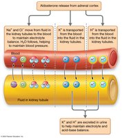
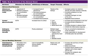
Mineralocorticoids (Aldosterone): Regulate sodium, potassium, and acid-base balance; increase blood pressure by promoting sodium and water retention.
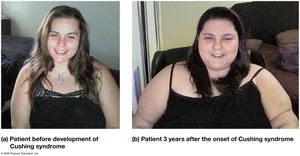
Glucocorticoids (Cortisol): Mediate stress response, increase blood glucose, suppress inflammation.
Androgenic steroids: Minor role in adults; contribute to development of secondary sex characteristics.
Hormones of the Adrenal Medulla
Catecholamines (Epinephrine and Norepinephrine): Mediate the fight-or-flight response; increase heart rate, blood pressure, and blood glucose.
Pancreas and Glucose Homeostasis
Structure of the Pancreas
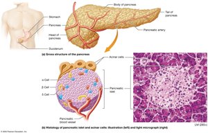
The pancreas contains both exocrine (acinar cells) and endocrine (islets of Langerhans) components. The islets contain alpha, beta, and delta cells that secrete glucagon, insulin, and somatostatin, respectively.
Hormones of the Endocrine Pancreas
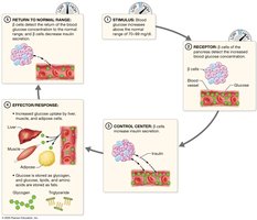
Glucagon: Increases blood glucose by promoting glycogen breakdown, gluconeogenesis, and fat breakdown.
Insulin: Lowers blood glucose by promoting glucose uptake and storage; stimulates glycogen and fat synthesis.
Somatostatin: Inhibits secretion of both insulin and glucagon.
Blood Glucose Regulation
Blood glucose is tightly regulated by the opposing actions of insulin and glucagon through negative feedback mechanisms.
Summary Tables
For a comprehensive overview, refer to the included tables summarizing the hormones, their stimuli for release, target tissues, and effects.
*Additional info: This summary covers the main content of Chapter 16 (The Endocrine System) from a Human Anatomy & Physiology textbook, including structure, function, regulation, and clinical relevance of the endocrine organs and their hormones. The notes are structured to facilitate exam preparation and understanding of endocrine physiology for college-level students.*
