 뒤로
뒤로The Endocrine System: Structure, Function, and Regulation
스터디 가이드 - 스마트 노트
 자료에 맞춘 맞춤형 노트, 핵심 정의, 예시, 맥락을 확장해 제공합니다.
Chapter 16: The Endocrine System
Endocrine System Overview
The endocrine system is one of the two major regulatory systems of the body, working alongside the nervous system to maintain homeostasis. It consists of glands and organs that secrete hormones directly into the bloodstream, allowing for widespread and long-lasting effects throughout the body.
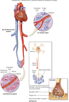
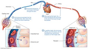
Endocrine glands are ductless and release hormones into the interstitial fluid, which then diffuse into blood capillaries.
Hormones are chemical messengers that regulate physiological processes such as growth, metabolism, and reproduction.
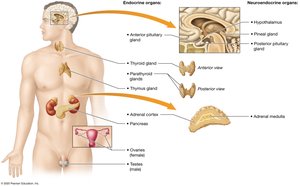
Endocrine System vs Nervous System
The endocrine and nervous systems both regulate body functions, but they differ in their mechanisms and effects:
The nervous system uses electrical impulses and neurotransmitters for rapid, short-term responses.
The endocrine system uses hormones transported by the blood for slower, but longer-lasting effects.
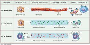
Paracrine and Autocrine Signals
Not all chemical signals act as classic hormones. Some act locally:
Paracrine signals: Chemicals secreted into the interstitial fluid that affect nearby cells without entering the bloodstream.
Autocrine signals: Chemicals secreted by cells that affect the same cell that secreted them.
Endocrine Organs
Endocrine organs are composed of specialized epithelial cells that secrete hormones into the interstitial fluid for transport via the bloodstream. This is in contrast to exocrine glands, which secrete substances through ducts to the exterior or into body cavities.
Primary Endocrine Organs
Pituitary gland
Thyroid gland
Parathyroid glands
Adrenal glands
Pineal gland
Secondary Endocrine Glands
Heart
Kidneys
Gastrointestinal tract
Thymus
Gonads (testes and ovaries)
Neuroendocrine Organs
Hypothalamus
Adrenal medulla
Classes of Hormones
Hormones are classified based on their chemical structure:
Amino acid-based hormones: Includes amines, peptides, and proteins; generally hydrophilic.
Steroid hormones: Derived from cholesterol; hydrophobic.
Hormone Transport
Hormones are transported in the blood, either freely (hydrophilic hormones) or bound to carrier proteins (hydrophobic hormones).
Target Cells and Receptors
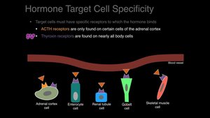
Hormones affect only specific target cells that possess the appropriate receptors. The specificity of hormone action is determined by the presence of these receptors.
Some hormones bind to only one type of target cell, while others can bind to multiple cell types.
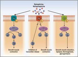
Binding to different receptors may produce opposite effects.
Location of Hormone Receptors
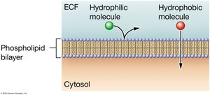
Receptors may be located on the plasma membrane (for hydrophilic hormones) or inside the cell (for hydrophobic hormones).
Regulation of Receptor Number
Upregulation: Increase in receptor number in response to low hormone levels.
Downregulation: Decrease in receptor number in response to high hormone levels.
Mechanism of Action
Hydrophilic Hormones
Hydrophilic hormones (amino acid-based and peptide/protein hormones) bind to receptors on the cell surface, initiating a cascade of events often involving second messengers such as cAMP.
Second-messenger systems amplify the hormone's signal within the cell.
Hydrophobic Hormones
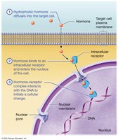
Hydrophobic hormones (steroids and thyroid hormones) diffuse through the plasma membrane and bind to intracellular receptors, often directly affecting gene expression.
Effects of Hormone Actions
Stimulate secretion from endocrine or exocrine cells
Activate or inhibit enzymes
Stimulate or inhibit mitosis and/or meiosis
Open or close ion channels, altering membrane potential
Activate or inhibit transcription of genes (gene expression)
Hormone Interactions
Hormones can interact in complex ways, including synergistic, antagonistic, and permissive effects.
Hormone Half-Life and Elimination
Hormones remain in the blood until taken up by target cells or broken down, primarily by the liver or kidneys. The half-life is the time required for half the hormone to be removed from the blood.
Regulation of Hormone Secretion
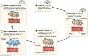
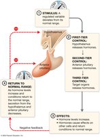
Hormone secretion is usually regulated by negative feedback loops involving a stimulus, receptor, control center, effector/response, and return to normal range.
Hypothalamus and Pituitary Gland
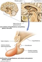
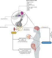
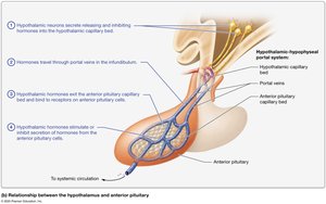
The hypothalamus is a key neuroendocrine organ that links the nervous and endocrine systems. It controls the pituitary gland via the infundibulum (pituitary stalk).
Anterior pituitary: Controlled by hypothalamic hormones via the hypophyseal portal system.
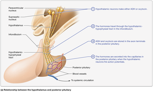
Posterior pituitary: Stores and releases hormones produced by the hypothalamus, transported via the hypothalamic-hypophyseal tract.
Posterior Pituitary Hormones
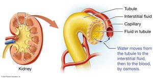
Antidiuretic hormone (ADH): Increases water retention by the kidneys, reducing urine output.
Oxytocin: Stimulates uterine contractions during childbirth and milk ejection during breastfeeding.
Anterior Pituitary Hormones
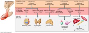
The anterior pituitary is regulated by releasing and inhibiting hormones from the hypothalamus. It secretes several tropic hormones that control other endocrine glands:
Thyroid-stimulating hormone (TSH): Stimulates thyroid gland.
Adrenocorticotropic hormone (ACTH): Stimulates adrenal cortex.
Prolactin (PRL): Stimulates milk production in mammary glands.
Luteinizing hormone (LH) and Follicle-stimulating hormone (FSH): Regulate gonadal function.
Growth hormone (GH): Stimulates growth and metabolism.
Growth Hormone (GH)
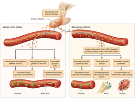
GH has both short-term metabolic effects and long-term effects on growth and development. It is regulated by GHRH and GHIH (somatostatin) from the hypothalamus.
Growth Hormone Disorders
Gigantism: Hypersecretion of GH before epiphyseal plate closure, leading to excessive growth.
Acromegaly: Hypersecretion of GH after epiphyseal plate closure, causing tissue overgrowth.
Pituitary dwarfism: Hyposecretion of GH, resulting in short stature with normal proportions.


Thyroid and Parathyroid Glands
Structure of the Thyroid Gland
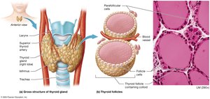
The thyroid gland is located in the anterior neck and consists of right and left lobes connected by an isthmus. It is composed of spherical follicles lined by follicular cells, which produce thyroid hormones. Parafollicular cells between follicles produce calcitonin.
Structure of the Parathyroid Glands
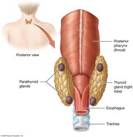
Usually four small glands embedded in the posterior surface of the thyroid. Chief cells produce parathyroid hormone (PTH).
Thyroid Hormones
Triiodothyronine (T3): Contains three iodine atoms; more active form.
Thyroxine (T4): Contains four iodine atoms; converted to T3 in target cells.
Regulate basal metabolic rate, thermoregulation, growth, and development.
Regulation of Thyroid Hormone Production
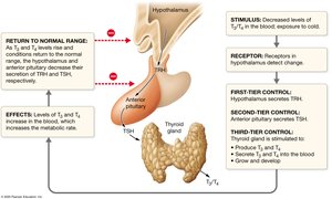
Controlled by a negative feedback loop involving TRH (hypothalamus), TSH (anterior pituitary), and T3/T4 (thyroid gland).
Thyroid Disorders
Hyperthyroidism: Excess thyroid hormone production (e.g., Graves disease).
Hypothyroidism: Insufficient thyroid hormone production (e.g., Hashimoto thyroiditis).
Parathyroid Hormone (PTH)
PTH is secreted in response to low blood calcium levels. It increases blood calcium by stimulating osteoclasts, increasing intestinal absorption, and enhancing renal reabsorption of calcium.
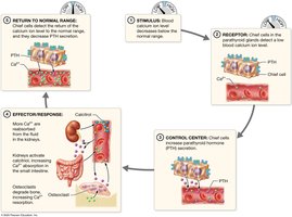
Calcitonin
Produced by parafollicular cells of the thyroid in response to high blood calcium. Inhibits osteoclast activity, lowering blood calcium levels.
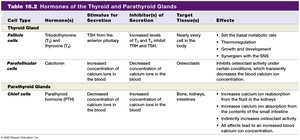
Summary Table: Hormones of the Thyroid and Parathyroid Glands
Cell Type | Hormone(s) | Stimulus for Secretion | Inhibitor(s) of Secretion | Target Tissue(s) | Effects |
|---|---|---|---|---|---|
Follicle cells | Thyroid hormones (T3, T4) | TSH from anterior pituitary | Increased T3/T4, somatostatin | Nearly every cell in the body | Set basal metabolic rate, thermoregulation, growth, development, synergism with SNS |
Parafollicular cells | Calcitonin | Increased blood calcium | Decreased blood calcium | Osteoclasts | Inhibits osteoclast activity, decreases blood calcium |
Chief cells (parathyroid) | PTH | Decreased blood calcium | Increased blood calcium | Bone, kidneys, intestine | Increases blood calcium by stimulating osteoclasts, increasing absorption and reabsorption |
Structure of the Adrenal Glands
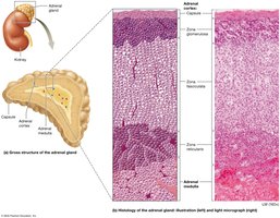
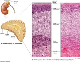
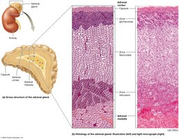
The adrenal glands are pyramid-shaped organs located on the superior aspect of each kidney. Each gland consists of an outer cortex and an inner medulla.
Adrenal Cortex
Zona glomerulosa: Produces mineralocorticoids (e.g., aldosterone).
Zona fasciculata: Produces glucocorticoids (e.g., cortisol).
Zona reticularis: Produces some glucocorticoids and androgenic steroids.
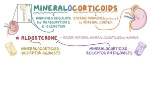
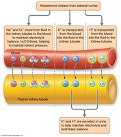
Mineralocorticoids
Regulate fluid and electrolyte balance. Aldosterone is the main mineralocorticoid, maintaining sodium and potassium levels, blood pressure, and acid-base homeostasis.
Glucocorticoids
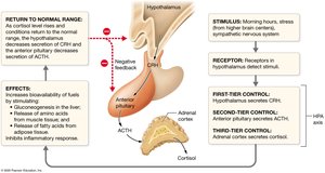
Regulate metabolism and the stress response. Cortisol is the most potent glucocorticoid, promoting gluconeogenesis, mobilizing amino acids and fatty acids, and suppressing inflammation.
Regulation of Cortisol Synthesis
Controlled by the HPA axis: CRH (hypothalamus) → ACTH (anterior pituitary) → cortisol (adrenal cortex).
Cortisol Disorders
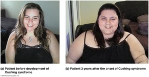
Cushing's Disease/Syndrome: Excess cortisol, leading to fat redistribution, muscle wasting, and immune suppression.
Androgenic Steroids
Produced in small amounts by the adrenal cortex; can be converted to testosterone or estrogen in circulation.
Adrenal Medulla
Composed of chromaffin cells derived from nervous tissue. Secretes epinephrine and norepinephrine in response to sympathetic stimulation, prolonging the fight-or-flight response.
Structure of the Pancreas
The pancreas is a club-shaped organ located posterior to the stomach. It contains both exocrine (acinar cells) and endocrine (islets of Langerhans) components.
Pancreatic Islets
Alpha cells: Secrete glucagon.
Beta cells: Secrete insulin.
Delta cells: Secrete somatostatin.
Glucagon
Increases blood glucose by promoting glycogenolysis, gluconeogenesis, and ketone body formation in the liver.
Insulin
Lowers blood glucose by promoting cellular uptake and storage of glucose, amino acids, and fats. Essential for glucose homeostasis.
Blood Glucose Regulation
Maintained by the opposing actions of insulin and glucagon in response to changes in blood glucose levels.
Diabetes Mellitus
Type 1: Autoimmune destruction of beta cells; requires insulin therapy.
Type 2: Insulin resistance and impaired beta cell function; associated with obesity and heredity.
The Thymus
Located in the mediastinum, the thymus is the site of T-lymphocyte maturation and secretes thymosin and thymopoietin.
The Gonads: Sex Hormones
Testes: Produce testosterone, regulated by LH and FSH.
Ovaries: Produce estrogens and progesterone, regulated by LH and FSH.
Pineal Gland
Located in the epithalamus, the pineal gland secretes melatonin, which regulates circadian rhythms.
Other Endocrine Tissues
Adipose tissue: Produces leptin, which regulates appetite.
Heart: Produces atrial natriuretic peptide (ANP), which lowers blood pressure.
Kidneys: Produce erythropoietin (EPO), renin, and activate vitamin D.
