 뒤로
뒤로The Endocrine System: Structure, Function, and Regulation
스터디 가이드 - 스마트 노트
 자료에 맞춘 맞춤형 노트, 핵심 정의, 예시, 맥락을 확장해 제공합니다.
The Endocrine System
Overview and Comparison with the Nervous System
The endocrine system is a major regulatory system of the body, working alongside the nervous system to maintain homeostasis. Unlike the nervous system, which uses electrical impulses and neurotransmitters for rapid, short-term responses, the endocrine system uses hormones released into the bloodstream for slower, longer-lasting effects.
Nervous system: Initiates rapid, short-duration responses via action potentials and neurotransmitters; acts at specific locations.
Endocrine system: Initiates slower, long-duration responses via hormones; acts at diffuse locations throughout the body.
Signal strength: Nervous system codes by frequency of action potentials; endocrine system by hormone concentration.
Major Endocrine Organs
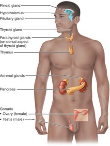
Endocrine organs are ductless glands that secrete hormones directly into the blood. Some organs are strictly endocrine, while others have both endocrine and exocrine functions.
Strictly endocrine: Pituitary, thyroid, pineal, parathyroid, adrenal glands
Endocrine + exocrine: Pancreas, gonads, thymus
Endocrine + neural: Hypothalamus
Hormone Action and Regulation
Hormone Types and Mechanisms
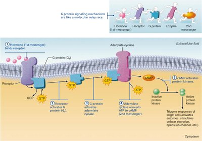
Hormones can be classified as amino acid-based (water-soluble) or steroid-based (lipid-soluble). Their solubility determines their mechanism of action and receptor location.
Amino acid-based hormones: Water-soluble, act on plasma membrane receptors, use second messenger systems (e.g., cAMP).
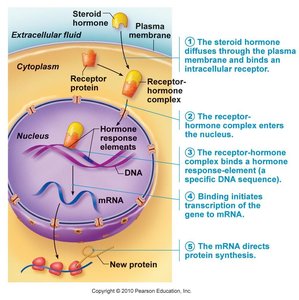
Steroid hormones: Lipid-soluble, diffuse across plasma membrane, act on intracellular receptors, directly activate genes.
Exception: Thyroid hormone is amino acid-based but acts like a steroid hormone.
Cyclic AMP (cAMP) Second Messenger Mechanism
Water-soluble hormones bind to receptors on the cell surface, activating G proteins, which then stimulate the production of cAMP. cAMP acts as a second messenger to activate protein kinases, leading to cellular responses.
Steroid Hormone Mechanism (Direct Gene Activation)
Steroid hormones diffuse through the plasma membrane and bind to intracellular receptors. The hormone-receptor complex enters the nucleus, binds to DNA, and initiates transcription of specific genes, resulting in protein synthesis.
Control of Hormone Release
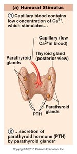
Hormone secretion is regulated by three main types of stimuli:
Humoral stimuli: Changes in blood levels of ions or nutrients (e.g., low Ca2+ stimulates parathyroid hormone release).
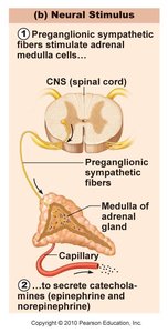
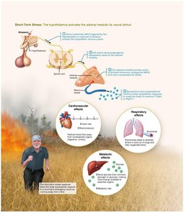
Neural stimuli: Nerve fibers stimulate hormone release (e.g., sympathetic stimulation of adrenal medulla).
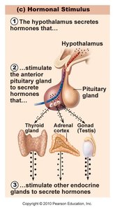
Hormonal stimuli: Hormones stimulate other endocrine glands to release hormones (e.g., hypothalamic hormones stimulate pituitary).
Hypothalamus and Pituitary Gland
Hypothalamus
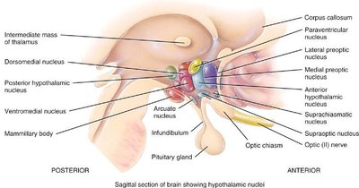
The hypothalamus is a key regulatory center that links the nervous and endocrine systems. It receives input from various brain regions and the body, and controls the pituitary gland via releasing and inhibiting hormones.
Pituitary Gland
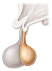
The pituitary gland is divided into anterior and posterior lobes, each releasing different hormones under hypothalamic control.
Posterior pituitary: Stores and releases oxytocin and antidiuretic hormone (ADH) produced by the hypothalamus.
Anterior pituitary: Produces hormones such as growth hormone (GH), thyroid-stimulating hormone (TSH), adrenocorticotropic hormone (ACTH), follicle-stimulating hormone (FSH), luteinizing hormone (LH), and prolactin (PRL).
Posterior Pituitary Hormones
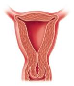
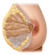
Oxytocin: Stimulates uterine contractions during labor and milk ejection during breastfeeding.
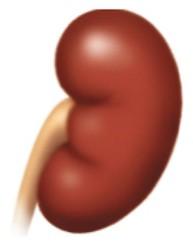
Antidiuretic hormone (ADH): Promotes water reabsorption in the kidneys; deficiency causes diabetes insipidus.
Anterior Pituitary Hormones
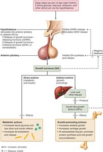
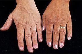
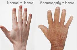
Growth hormone (GH): Stimulates growth and metabolism; excess causes gigantism/acromegaly, deficiency causes dwarfism.
Thyroid-stimulating hormone (TSH): Stimulates thyroid hormone release.
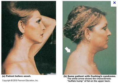
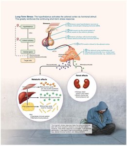
Adrenocorticotropic hormone (ACTH): Stimulates adrenal cortex to release glucocorticoids.
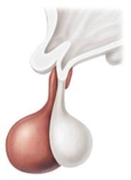
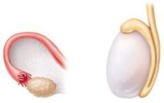
Follicle-stimulating hormone (FSH) and Luteinizing hormone (LH): Regulate gonadal function and hormone production.
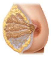
Prolactin (PRL): Promotes lactation.
Growth Hormone Disorders
Acromegaly: Excess GH in adults, causing enlarged extremities.
Gigantism: Excess GH in children, causing abnormal height.
Dwarfism: GH deficiency in children, causing short stature.

Thyroid and Parathyroid Glands
Thyroid Gland
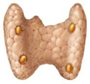
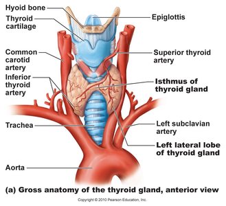
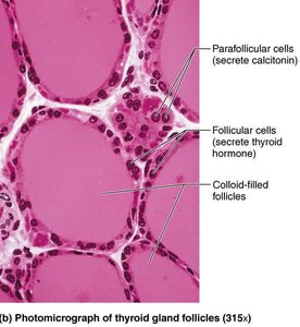
The thyroid gland produces thyroid hormones (T3 and T4) that regulate metabolism, growth, and development. It also produces calcitonin, which lowers blood calcium levels.

Regulation of Thyroid Hormone Secretion
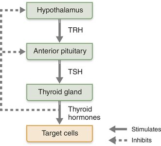
Thyroid hormone release is regulated by the hypothalamic-pituitary-thyroid axis:
Hypothalamus releases TRH (thyrotropin-releasing hormone)
TRH stimulates anterior pituitary to release TSH
TSH stimulates thyroid gland to release T3 and T4
Negative feedback from thyroid hormones inhibits TRH and TSH release
Thyroid Hormone Synthesis
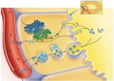
Thyroid hormone synthesis involves trapping iodide, oxidizing it to iodine, and attaching it to tyrosine residues in thyroglobulin. The iodinated tyrosines are coupled to form T3 and T4, which are released into the bloodstream.
Thyroid Pathology
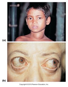
Goiter: Enlargement of the thyroid gland, often due to iodine deficiency.
Hashimoto’s disease: Autoimmune hypothyroidism.
Myxedema: Severe hypothyroidism in adults.
Graves’ disease: Autoimmune hyperthyroidism, often with exophthalmos (protruding eyes).
Parathyroid Glands
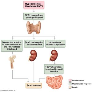
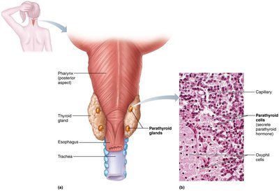
The parathyroid glands secrete parathyroid hormone (PTH), which increases blood calcium levels by stimulating osteoclasts, increasing calcium reabsorption in the kidneys, and activating vitamin D.
Adrenal Glands
Structure and Hormones
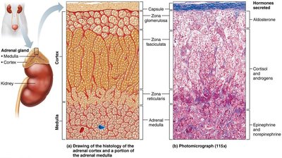
The adrenal glands consist of the cortex (producing corticosteroids) and the medulla (producing catecholamines).
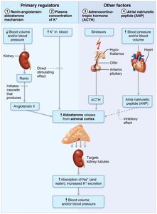
Mineralocorticoids (aldosterone): Regulate sodium and potassium balance.
Glucocorticoids (cortisol): Regulate metabolism and stress response.
Gonadocorticoids (androgens): Contribute to sex characteristics.
Catecholamines (epinephrine, norepinephrine): Mediate fight-or-flight response.
Pancreas
Endocrine and Exocrine Functions
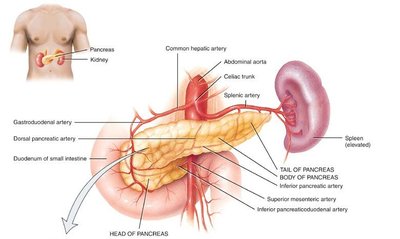
The pancreas has both endocrine (islets of Langerhans) and exocrine (acinar cells) functions. The endocrine portion regulates blood glucose via insulin (lowers glucose) and glucagon (raises glucose).
Other Endocrine Organs and Hormones
Pineal gland: Secretes melatonin, regulates circadian rhythms.
Adipose tissue: Leptin regulates appetite and metabolism.
Heart: Atrial natriuretic peptide (ANP) lowers blood pressure.
Kidney: Erythropoietin (EPO) stimulates red blood cell production.
Skin: Produces cholecalciferol (vitamin D3 precursor).
Thymus: Produces hormones for T lymphocyte development.
Summary Table: Comparison of Lipid- and Water-Soluble Hormones
Lipid-soluble hormones | Water-soluble hormones | |
|---|---|---|
Consist of | All steroid hormones and thyroid hormone | All amino acid–based hormones except thyroid hormone |
Sources | Adrenal cortex, gonads, thyroid gland | All other endocrine glands |
Stored in secretory vesicles | No | Yes |
Transport in blood | Bound to plasma proteins | Usually free in plasma |
Half-life in blood | Long (metabolized by liver) | Short (removed by kidneys) |
Location of receptors | Usually inside cell | On plasma membrane |
Mechanism of action | Activate genes, cause new protein synthesis | Act through second-messenger systems |
