 뒤로
뒤로The Endocrine System: Structure, Function, and Regulation
스터디 가이드 - 스마트 노트
 자료에 맞춘 맞춤형 노트, 핵심 정의, 예시, 맥락을 확장해 제공합니다.
The Endocrine System
Overview of the Endocrine System
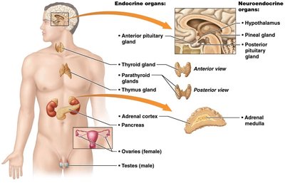
The endocrine system works in conjunction with the nervous system to coordinate and integrate the activity of body cells. It influences metabolic activities by means of hormones transported in the blood. Endocrine responses are generally slower but longer-lasting than those of the nervous system. The study of hormones and endocrine organs is known as endocrinology.
Major functions: Reproduction, growth and development, maintenance of electrolyte, water, and nutrient balance, regulation of cellular metabolism and energy balance, and mobilization of body defenses.
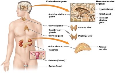
Endocrine glands: Pituitary, thyroid, parathyroid, adrenal, and pineal glands.
Neuroendocrine organ: Hypothalamus.
Other hormone-producing tissues: Pancreas, gonads, placenta, adipose cells, thymus, and cells in the small intestine, stomach, kidneys, and heart.
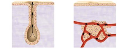
Exocrine vs. Endocrine Glands
Exocrine glands produce nonhormonal substances (e.g., sweat, saliva) and have ducts to carry secretions to membrane surfaces. Endocrine glands produce hormones and lack ducts, releasing their products directly into the bloodstream.
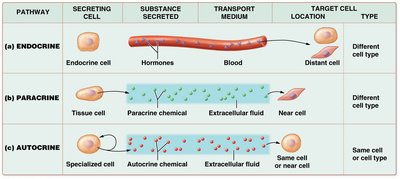
Chemical Messengers of the Endocrine System
The endocrine system uses several types of chemical messengers:
Hormones: Long-distance chemical signals that travel in blood or lymph.
Autocrines: Chemicals that exert effects on the same cells that secrete them (not considered hormones).
Paracrines: Locally acting chemicals that affect cells other than those that secrete them (not considered hormones).
Classes of Hormones
Hormones are classified into two main groups based on their chemical structure:
Amino acid–based hormones: Includes amino acid derivatives, peptides, and proteins. Most hormones belong to this class.
Steroid hormones: Synthesized from cholesterol; includes gonadal and adrenocortical hormones. These can cross cell membranes and directly affect DNA transcription.
Hormone Action and Target Cells
Although hormones circulate systemically, only cells with specific receptors (target cells) are affected. Hormones alter target cell activity by:
Altering plasma membrane permeability or membrane potential
Stimulating synthesis of enzymes or other proteins
Activating or deactivating enzymes
Inducing secretory activity
Stimulating mitosis
Mechanisms of Hormone Action
Hormones act in one of two ways, depending on their chemical nature and receptor location:
Water-soluble hormones (all amino acid–based hormones except thyroid hormone): Act on plasma membrane receptors via G protein second messengers; cannot enter the cell.
Lipid-soluble hormones (steroid and thyroid hormones): Act on intracellular receptors that directly activate genes; can enter the cell.
Second Messenger Systems
Water-soluble hormones often use the cyclic AMP (cAMP) second-messenger mechanism:
Hormone (first messenger) binds to receptor.
Receptor activates G protein.
G protein activates adenylate cyclase.
Adenylate cyclase converts ATP to cAMP (second messenger).
cAMP activates protein kinases, which trigger cellular responses.
Direct Gene Activation
Lipid-soluble hormones diffuse through the plasma membrane, bind to intracellular receptors, and directly activate gene transcription, leading to protein synthesis.
Regulation of Hormone Release
Endocrine Gland Stimuli
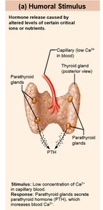
Endocrine glands are stimulated to synthesize and release hormones in response to three types of stimuli:
Humoral stimuli: Changing blood levels of ions and nutrients directly stimulate hormone secretion (e.g., low Ca2+ stimulates parathyroid hormone release).
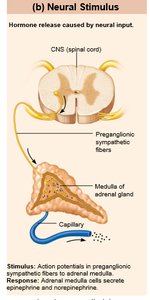
Neural stimuli: Nerve fibers stimulate hormone release (e.g., sympathetic nervous system stimulates adrenal medulla to secrete catecholamines).
Hormonal stimuli: Hormones stimulate other endocrine organs to release their hormones (e.g., hypothalamic hormones stimulate anterior pituitary hormones).
The Hypothalamus and Pituitary Gland
Structure and Function
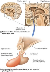
The hypothalamus is connected to the pituitary gland (hypophysis) via the infundibulum. The pituitary gland has two major lobes:
Posterior pituitary (neurohypophysis): Composed of neural tissue; stores and releases neurohormones (oxytocin and antidiuretic hormone, ADH).
Anterior pituitary (adenohypophysis): Consists of glandular tissue; produces and releases several hormones under hypothalamic control.
Hormone Release Mechanisms
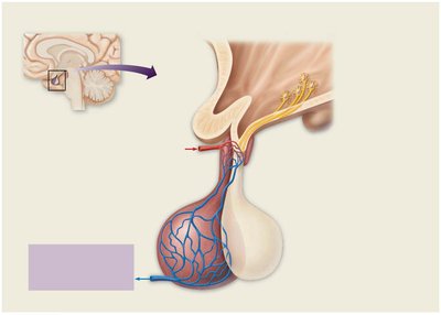
Posterior pituitary: Hypothalamic neurons synthesize oxytocin and ADH, which are transported down axons and released into the blood from the posterior pituitary.
Anterior pituitary: Hypothalamic hormones are released into the hypophyseal portal system, stimulating or inhibiting the release of anterior pituitary hormones.
Major Pituitary Hormones
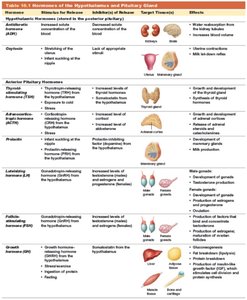
Oxytocin: Stimulates uterine contractions and milk ejection; acts via positive feedback.
Antidiuretic hormone (ADH): Promotes water reabsorption in kidneys; inhibited by alcohol and diuretics.
Growth hormone (GH): Stimulates growth, protein synthesis, and mobilizes fat; regulated by GHRH and GHIH.
Thyroid-stimulating hormone (TSH): Stimulates thyroid gland activity.
Adrenocorticotropic hormone (ACTH): Stimulates adrenal cortex to release corticosteroids.
Follicle-stimulating hormone (FSH) and Luteinizing hormone (LH): Regulate function of gonads.
Prolactin (PRL): Stimulates milk production.
Growth Hormone Disorders
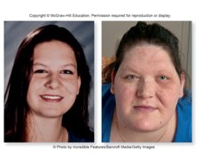
Hypersecretion: Causes gigantism in children and acromegaly in adults.
Hyposecretion: Causes pituitary dwarfism in children.

The Thyroid and Parathyroid Glands
Thyroid Gland
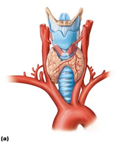
The thyroid gland is located in the anterior neck and produces thyroid hormones (T3 and T4), which regulate metabolism, and calcitonin, which helps regulate calcium levels.
Parathyroid Glands
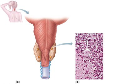
The parathyroid glands are small glands on the posterior aspect of the thyroid. They secrete parathyroid hormone (PTH), the most important hormone in calcium homeostasis.
Stimulates osteoclasts to release Ca2+ from bone
Enhances reabsorption of Ca2+ by kidneys
Promotes activation of vitamin D for increased intestinal absorption of Ca2+
The Adrenal Glands
Structure and Hormones
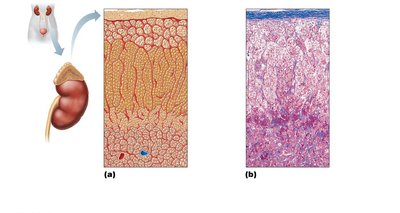
The adrenal glands are paired organs atop the kidneys, consisting of the adrenal cortex and adrenal medulla.
Adrenal cortex: Produces corticosteroids in three layers:
Zona glomerulosa: Mineralocorticoids (e.g., aldosterone)
Zona fasciculata: Glucocorticoids (e.g., cortisol)
Zona reticularis: Gonadocorticoids (sex hormones)
Adrenal medulla: Produces catecholamines (epinephrine and norepinephrine) for the fight-or-flight response.
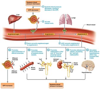
Regulation of Aldosterone
Aldosterone secretion is regulated by:
Renin-angiotensin-aldosterone mechanism
Plasma concentration of K+
ACTH
Atrial natriuretic peptide (ANP)
Adrenal Medulla and Stress Response
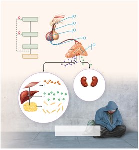
The adrenal medulla releases catecholamines in response to stress, increasing heart rate, blood pressure, and blood glucose, and diverting blood to essential organs.
The Pineal Gland
Structure and Function
The pineal gland is a small gland hanging from the roof of the third ventricle. It secretes melatonin, which regulates sleep-wake cycles and may influence puberty timing.
The Pancreas
Structure and Function
The pancreas has both exocrine (digestive enzyme production) and endocrine (hormone production) functions. The endocrine portion consists of pancreatic islets:
Alpha cells: Produce glucagon (raises blood glucose)
Beta cells: Produce insulin (lowers blood glucose)
Regulation of Blood Glucose
Glucagon and insulin have opposing effects to maintain blood glucose homeostasis:
Glucagon: Stimulates glycogen breakdown and gluconeogenesis in the liver, raising blood glucose.
Insulin: Enhances glucose uptake by cells, inhibits glycogen breakdown, and promotes glucose storage as glycogen or fat.
The Gonads and Placenta
Hormones and Functions
Ovaries: Produce estrogens and progesterone, regulating reproductive organ maturation, secondary sexual characteristics, and menstrual cycle.
Testes: Produce testosterone, responsible for male reproductive organ maturation, secondary sexual characteristics, and sperm production.
Placenta: Secretes estrogens, progesterone, and human chorionic gonadotropin (hCG) during pregnancy.
Hormone Secretion by Other Organs
Adipose tissue: Leptin (appetite control), resistin (insulin antagonist), adiponectin (enhances insulin sensitivity)
Gastrointestinal tract: Gastrin, ghrelin, secretin, cholecystokinin (CCK), incretins
Heart: Atrial natriuretic peptide (ANP) lowers blood pressure
Kidneys: Erythropoietin (stimulates red blood cell production), renin (regulates blood pressure)
Skeleton: Osteocalcin (regulates insulin secretion and sensitivity)
Skin: Cholecalciferol (vitamin D precursor), calcitriol (active vitamin D)
Thymus: Thymulin, thymopoietins, thymosins (T cell development)
