 뒤로
뒤로The Heart: Structure, Function, and Physiology
스터디 가이드 - 스마트 노트
 자료에 맞춘 맞춤형 노트, 핵심 정의, 예시, 맥락을 확장해 제공합니다.
The Heart: Structure, Function, and Physiology
An Introduction to the Heart
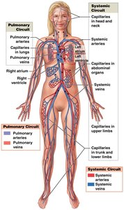
The heart is the central organ of the cardiovascular system, responsible for pumping blood throughout the body. It beats approximately 100,000 times per day, moving about 8,000 liters of blood. The cardiovascular system consists of the heart, blood, and blood vessels, which together maintain the circulation of blood and the delivery of oxygen and nutrients to tissues.
Pulmonary circuit: Carries blood to and from the lungs for gas exchange.
Systemic circuit: Delivers blood to and from the rest of the body.
Blood flows through these circuits in sequence, beginning and ending at the heart.
Anatomy of the Heart
Types of Blood Vessels
Arteries: Carry blood away from the heart.
Veins: Return blood to the heart.
Capillaries: Smallest vessels where exchange of gases, nutrients, and wastes occurs between blood and tissues.
Chambers of the Heart
Right atrium: Receives blood from the systemic circuit.
Right ventricle: Pumps blood into the pulmonary circuit.
Left atrium: Receives blood from the pulmonary circuit.
Left ventricle: Pumps blood into the systemic circuit.
Location and Orientation
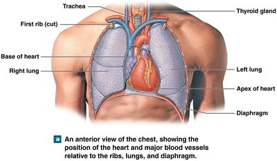
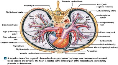
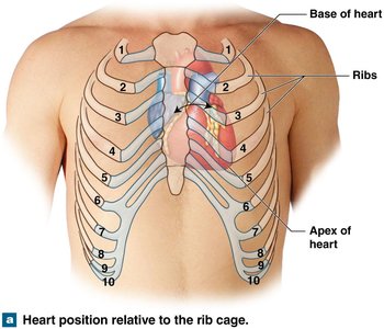
The heart is located in the mediastinum, between the two pleural cavities, with its base superior and apex inferior. Major vessels connect at the base, and the apex points downward and to the left.
Pericardium
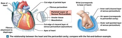
Fibrous pericardium: Outer tough layer.
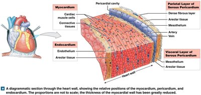
Serous pericardium: Inner double-layered membrane (parietal and visceral layers).
Pericardial cavity: Space between parietal and visceral layers, containing pericardial fluid to reduce friction.
Superficial Anatomy
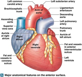
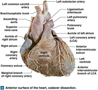
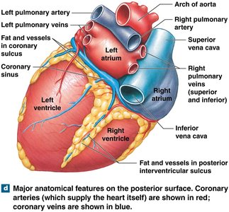
Two thin-walled atria, each with an expandable auricle.
Sulci (grooves) on the heart surface contain fat and blood vessels.
Coronary sulcus: Separates atria from ventricles.
Anterior and posterior interventricular sulci: Mark the boundary between left and right ventricles.
Heart Wall Structure
Epicardium: Outer visceral layer of serous pericardium.
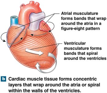
Myocardium: Middle layer of cardiac muscle tissue, responsible for contraction.
Endocardium: Inner layer of simple squamous epithelium and areolar tissue.
Connective Tissues and Cardiac Skeleton
Support cardiac muscle fibers, blood vessels, and nerves.
Distribute contraction forces and add strength.
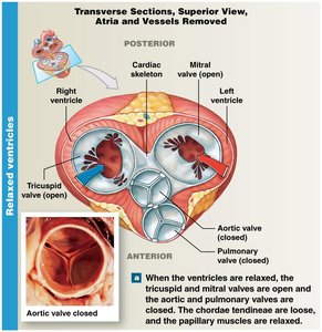
Cardiac skeleton: Four dense bands of elastic tissue encircle valves and bases of major vessels, stabilizing and electrically insulating the heart.
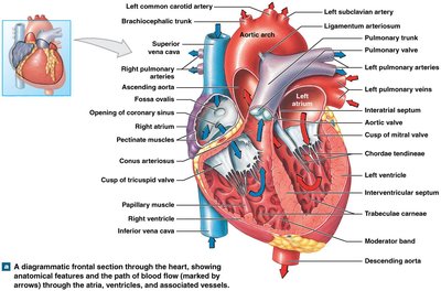
Internal Anatomy and Valves
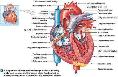
Chambers separated by septa: Interatrial septum (between atria), interventricular septum (between ventricles, thicker).
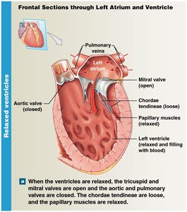
Atrioventricular (AV) valves: Tricuspid (right) and mitral (left) valves, prevent backflow into atria.
Semilunar valves: Pulmonary and aortic valves, prevent backflow into ventricles.
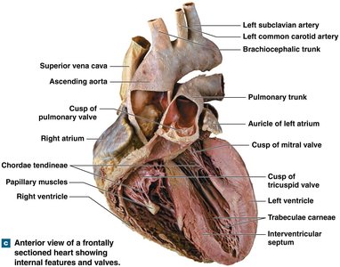
Right and Left Heart Structures
Right atrium: Receives blood from superior and inferior vena cava; contains pectinate muscles and fossa ovalis (remnant of fetal foramen ovale).
Right ventricle: Contains trabeculae carneae and moderator band; pumps blood to pulmonary trunk via pulmonary valve.
Left atrium: Receives blood from pulmonary veins; passes blood to left ventricle through mitral valve.
Left ventricle: Thicker wall, pumps blood to systemic circuit via aortic valve; forms aortic arch and descending aorta.
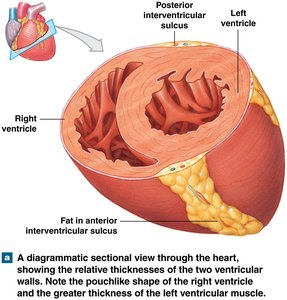
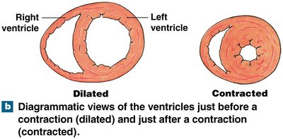
Ventricular Structure Comparison
Both ventricles pump equal volumes of blood.
Right ventricle: Thinner wall, lower pressure, pouch-shaped.
Left ventricle: Thicker wall, higher pressure, round shape.
Heart Valves and Blood Flow
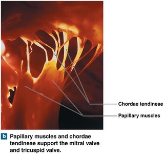
AV valves prevent backflow into atria during ventricular contraction; chordae tendineae and papillary muscles prevent valve inversion.
Semilunar valves prevent backflow into ventricles; no muscular support.
Valvular heart disease (VHD) may result from inflammation or rheumatic fever.
Coronary Circulation
Coronary circulation supplies blood to the heart muscle itself. Coronary arteries originate at the aortic sinuses and maintain blood flow through elastic rebound of the aorta.
Right coronary artery: Supplies right atrium, portions of both ventricles, and conduction system; branches into marginal and posterior interventricular arteries.
Left coronary artery: Supplies left ventricle, left atrium, and interventricular septum; branches into circumflex and anterior interventricular arteries.
Cardiac veins: Great cardiac vein, middle cardiac vein, small cardiac vein, and coronary sinus return deoxygenated blood to the right atrium.
Clinical Correlations: Coronary Artery Disease and Myocardial Infarction
Coronary artery disease (CAD): Partial or complete blockage of coronary arteries, usually due to atherosclerotic plaque or thrombus.
Angina pectoris: Chest pain due to temporary ischemia during increased workload.
Myocardial infarction (MI): Heart attack caused by prolonged blockage, leading to death of cardiac muscle cells and formation of nonfunctional tissue (infarct).
Diagnosis: ECG and blood tests for cardiac enzymes (troponin T, troponin I, CK-MB).
Treatments: Risk factor modification, drug therapy (anticoagulants, beta-blockers, vasodilators, calcium channel blockers), noninvasive surgery (atherectomy, balloon angioplasty, stents), and coronary artery bypass graft (CABG).
The Conducting System of the Heart
The heart's conducting system consists of specialized cardiac muscle cells that initiate and distribute electrical impulses, ensuring coordinated contraction (autorhythmicity).
Pacemaker cells: Located in the sinoatrial (SA) node (right atrium) and atrioventricular (AV) node (junction of atria and ventricles).
Conducting cells: Found in internodal pathways, AV bundle, bundle branches, and Purkinje fibers.
SA node sets the sinus rhythm (60–100 action potentials/min); AV node (40–60/min).
Impulse Conduction Pathway
SA node activity and atrial activation begin.
Stimulus spreads across atria to AV node.
Impulse delayed at AV node; atrial contraction begins.
Impulse travels through AV bundle, bundle branches, Purkinje fibers, and moderator band to papillary muscles.
Impulse distributed to ventricular myocardium; ventricular contraction begins.
Electrocardiogram (ECG)
Records electrical events in the heart using electrodes on the body surface.
P wave: Atrial depolarization.
QRS complex: Ventricular depolarization (ventricles contract).
T wave: Ventricular repolarization.
Intervals (P–R, Q–T) are clinically important for diagnosing arrhythmias.
Cardiac Muscle Physiology
Cardiac contractile cells form the bulk of the heart wall and are interconnected by intercalated discs (desmosomes and gap junctions).
Action potential phases: rapid depolarization (Na+ influx), plateau (Ca2+ influx), repolarization (K+ efflux).
Refractory period prevents tetany, ensuring rhythmic contractions.
Calcium ions are essential for contraction; both extracellular and sarcoplasmic reticulum sources are involved.
Cardiac muscle relies on aerobic metabolism (fatty acids, glucose, myoglobin stores oxygen).
The Cardiac Cycle
The cardiac cycle is the sequence of events from the start of one heartbeat to the next, including alternating periods of contraction (systole) and relaxation (diastole).
Atrial systole: Atria contract, pushing blood into ventricles.
Ventricular systole: Ventricles contract, ejecting blood into arteries.
Diastole: Chambers relax and fill with blood.
Blood flows from high to low pressure, controlled by valve function.
Phases of the Cardiac Cycle
Atrial contraction (systole) forces additional blood into relaxed ventricles.
Ventricular contraction (systole) closes AV valves (isovolumetric contraction), then opens semilunar valves for ejection (stroke volume).
Ventricular relaxation (diastole) closes semilunar valves, ventricles fill passively.
Heart Sounds
S1: AV valves close ("lubb").
S2: Semilunar valves close ("dupp").
S3, S4: Soft sounds from blood flow and atrial contraction.
Heart murmur: Abnormal sound from valve regurgitation.
Cardiac Output
Cardiac output (CO) is the volume of blood pumped by the left ventricle per minute. It is a key indicator of heart function and tissue perfusion.
Formula:
Stroke volume (SV):
End-diastolic volume (EDV): Blood in ventricle at end of diastole.
End-systolic volume (ESV): Blood remaining after systole.
Ejection fraction: Percentage of EDV ejected per beat.
Factors Affecting Cardiac Output
Heart rate (HR): Influenced by autonomic nervous system, hormones, venous return, and reflexes (e.g., Bainbridge reflex).
Stroke volume (SV): Affected by preload (ventricular stretch), contractility (force of contraction), and afterload (resistance to ejection).
Frank–Starling Principle: As EDV increases, SV increases (within physiological limits).
Factor | Effect on Cardiac Output |
|---|---|
Increased HR | Increases CO (up to a point) |
Increased SV | Increases CO |
Increased preload | Increases SV and CO |
Increased contractility | Increases SV and CO |
Increased afterload | Decreases SV and CO |
Autonomic Regulation
Sympathetic stimulation (NE, E): Increases HR and contractility.
Parasympathetic stimulation (ACh): Decreases HR.
Hormones (epinephrine, norepinephrine, thyroid hormone): Increase HR and contractility.
Cardiac Reserve
Difference between resting and maximal cardiac output.
Indicates the heart's ability to respond to increased demands.
Additional info: This summary integrates and expands upon the provided lecture slides and textbook images, ensuring a comprehensive, exam-ready overview of the heart's anatomy and physiology for college-level Anatomy & Physiology students.
