 뒤로
뒤로The Lymphatic System and Immunity: Structure, Function, and Clinical Relevance
스터디 가이드 - 스마트 노트
 자료에 맞춘 맞춤형 노트, 핵심 정의, 예시, 맥락을 확장해 제공합니다.
The Lymphatic System and Immunity
Overview of the Lymphatic System
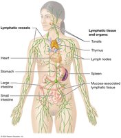
The lymphatic system is a network of organs, tissues, and vessels that collaborates with the immune system to defend the body against pathogens and maintain fluid balance. It consists of lymphatic vessels, lymphoid tissues, and organs such as lymph nodes, spleen, and thymus.
Lymphatic Vessels: Blind-ended vessels that collect interstitial fluid (lymph) and return it to the cardiovascular system.
Lymphoid Tissues and Organs: Include lymphoid follicles (e.g., tonsils), lymph nodes, spleen, and thymus.
Functions of the Lymphatic System
Regulation of Interstitial Fluid Volume: Collects excess interstitial fluid and returns it to the bloodstream, preventing edema.
Absorption of Dietary Fats: Specialized lymphatic vessels (lacteals) in the small intestine absorb fats and deliver them to the blood.
Immune Functions: Lymphoid organs filter pathogens and house leukocytes for immune surveillance.
Lymphatic Vessels and Circulation
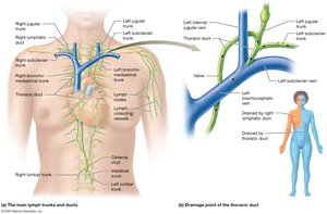
Lymphatic vessels collect lymph from tissues and transport it through progressively larger vessels and trunks, eventually draining into the venous system.
Lymph Trunks: Major trunks include lumbar, intestinal, jugular, bronchomediastinal, and subclavian trunks.
Cisterna Chyli: A dilated sac that receives lymph from the lower body and intestines.
Thoracic Duct: Drains lymph from most of the body into the left subclavian vein.
Right Lymphatic Duct: Drains lymph from the right upper body into the right subclavian vein.
Lymph Flow Mechanisms
No Central Pump: Lymph is propelled by skeletal muscle contractions, smooth muscle in vessel walls, and lymphatic valves that prevent backflow.
Lymphedema
Lymphedema is a severe, often disfiguring swelling caused by the accumulation of lymph due to vessel blockage or removal, commonly seen after lymph node dissection in cancer surgery.

Lymphatic Capillaries
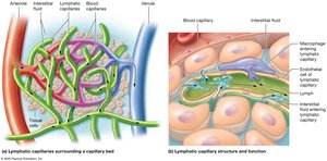
Structure: Blind-ended, highly permeable capillaries with overlapping endothelial cells that open in response to increased interstitial pressure.
Function: Allow entry of fluid, proteins, pathogens, and cells (e.g., macrophages, bacteria, cancer cells).
Lymphoid Tissues and Organs
Reticular Tissue
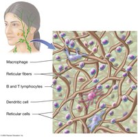
Reticular tissue forms the framework of lymphoid organs, consisting of reticular fibers that trap pathogens and support immune cells.
Lymphocytes: B cells and T cells.
Phagocytes: Macrophages and dendritic cells.
Reticular Cells: Produce reticular fibers.
Mucosa-Associated Lymphatic Tissue (MALT)
MALT consists of clusters of lymphoid tissue that protect mucous membranes exposed to pathogens, found in the gastrointestinal, respiratory, and genitourinary tracts.
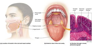
Tonsils: Pharyngeal, palatine, and lingual tonsils trap pathogens entering through the oral and nasal cavities.
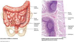
Peyer’s Patches: Aggregated lymphoid nodules in the ileum of the small intestine.
Appendix: Contains lymphoid tissue to defend against bacteria in the large intestine.
Lymph Nodes
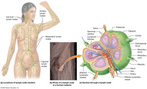
Lymph nodes are small, bean-shaped structures that filter lymph and house immune cells. They are found in clusters in the axillae, neck, groin, and abdomen.
Structure: Outer cortex (B cell-rich follicles), inner medulla (macrophages, mature B cells), and trabeculae.
Function: Trap pathogens and initiate immune responses.
Spleen
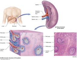
The spleen is the largest lymphoid organ, filtering blood and removing old erythrocytes. It contains red pulp (macrophages) and white pulp (lymphocytes).
Thymus
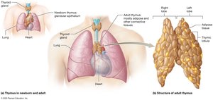
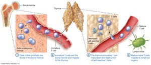
The thymus is the site of T cell maturation, most active in childhood and replaced by fat in adulthood. It ensures self-tolerance and immunocompetence of T cells.
Overview of the Immune System
Lines of Defense
First Line: Surface barriers (skin and mucous membranes).
Second Line: Innate immunity (cells and proteins that respond nonspecifically).
Third Line: Adaptive immunity (specific responses by T and B cells).
Categories of Immunity
Innate Immunity: Nonspecific, rapid response to all pathogens; includes phagocytes, natural killer cells, and complement proteins.
Adaptive Immunity: Specific, slower response involving T cells (cell-mediated) and B cells (antibody-mediated); characterized by immunological memory.
Surface Barriers
Skin: Multiple layers of keratinized epithelium resist mechanical stress and pathogen entry.
Mucous Membranes: Line body passages, secrete mucus to trap pathogens, and contain cilia to expel debris.
Secretions: Sebum, acid, defensins, and normal flora inhibit pathogen growth.
Cells and Proteins of the Immune System
Cells
Agranulocytes: B and T lymphocytes, monocytes (which become macrophages).
Granulocytes: Neutrophils, eosinophils, basophils.
Phagocytes: Engulf and destroy pathogens (macrophages, neutrophils, dendritic cells).
Natural Killer (NK) Cells: Destroy infected or cancerous cells without antigen specificity.
Proteins
Antibodies: Produced by B cells, bind specific antigens.
Complement System: Plasma proteins that enhance phagocytosis, inflammation, and cell lysis.
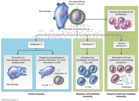
Cytokines: Signaling proteins that regulate immune responses (e.g., interleukins, interferons, tumor necrosis factor).
Innate Immunity: Mechanisms and Responses
Phagocytosis
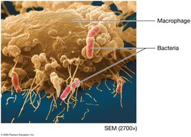
Macrophages: Engulf pathogens, present antigens, and secrete cytokines.
Neutrophils: Rapid responders, destroy bacteria via respiratory burst.
Eosinophils: Target parasitic pathogens.
Dendritic Cells: Present antigens to T and B cells, initiating adaptive responses.
Complement System
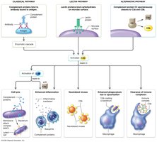
The complement system is activated via three pathways (classical, lectin, alternative) and leads to cell lysis, enhanced inflammation, opsonization, and clearance of immune complexes.
Inflammatory Response
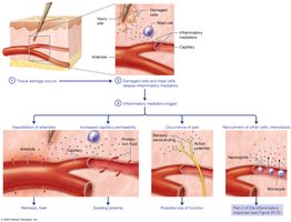
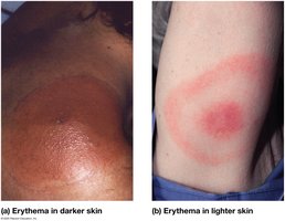
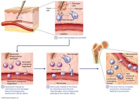
Inflammation is a protective response to tissue injury or infection, characterized by redness, heat, swelling, pain, and loss of function. It involves the release of mediators (histamine, cytokines), increased blood flow, and recruitment of phagocytes.
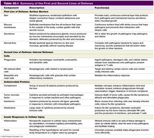
Summary Table: First and Second Lines of Defense
Component | Description | Function(s) |
|---|---|---|
Skin | Stratified, keratinized epithelium | Physical barrier, resists mechanical stress |
Mucous Membranes | Epithelium lining body passages | Traps pathogens, cilia expel debris |
Phagocytes | Macrophages, neutrophils, dendritic cells | Engulf and destroy pathogens |
Complement Proteins | Plasma proteins | Opsonization, cell lysis, inflammation |
Cytokines | Signaling proteins | Regulate immune cell activity |
Fever | Elevated body temperature | Enhances phagocyte function |
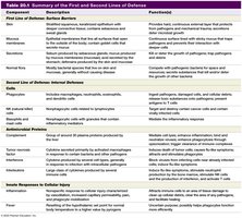
Additional info: | See images for full table |
Adaptive Immunity: Cell-Mediated and Antibody-Mediated Responses
T Cells and Cell-Mediated Immunity
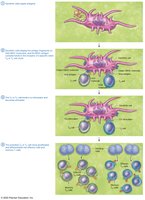
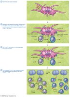
Helper T Cells (CD4+): Activate other immune cells via cytokine secretion.
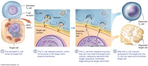
Cytotoxic T Cells (CD8+): Destroy infected or abnormal cells by inducing apoptosis.
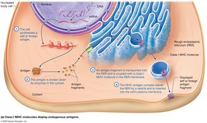
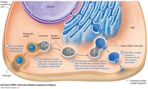
Major Histocompatibility Complex (MHC): MHC I presents endogenous antigens to CD8+ T cells; MHC II presents exogenous antigens to CD4+ T cells.
B Cells and Antibody-Mediated Immunity
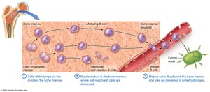
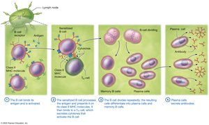
B Cell Activation: B cells bind antigen, present it to helper T cells, and differentiate into plasma cells (secrete antibodies) and memory B cells.
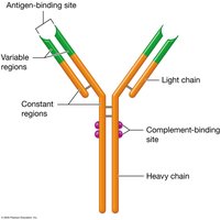
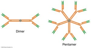
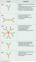
Antibody Structure: Y-shaped molecules with variable (antigen-binding) and constant regions; can form monomers, dimers, or pentamers.
Antibody Classes: IgG, IgA, IgM, IgE, IgD (mnemonic: GAMED).
Primary and Secondary Immune Responses
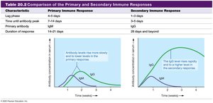
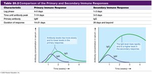
Primary Response: First exposure, slower antibody production (mainly IgM).
Secondary Response: Faster, stronger response (mainly IgG) due to memory cells.
Active and Passive Immunity
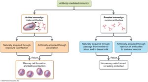
Active Immunity: Body produces its own antibodies (infection or vaccination); long-lasting.
Passive Immunity: Preformed antibodies are transferred (e.g., maternal IgG, antivenins); short-lived.
Disorders of the Immune System
Hypersensitivity Disorders
Type I (Immediate): Allergies mediated by IgE and mast cells (e.g., hay fever, asthma, anaphylaxis).
Type II (Antibody-Mediated): Antibodies target self-antigens (e.g., transfusion reactions).
Type III (Immune Complex-Mediated): Immune complexes deposit in tissues, causing inflammation (e.g., lupus).
Type IV (Delayed-Type): T cell-mediated (e.g., contact dermatitis, tuberculosis test).
Immunodeficiency Disorders
Primary: Genetic defects affecting immune components (e.g., SCID).
Secondary: Acquired due to infection (e.g., HIV/AIDS), cancer, or immunosuppressive therapy.
Autoimmune Disorders
Immune system attacks self-antigens, leading to tissue damage (e.g., multiple sclerosis, type 1 diabetes, systemic lupus erythematosus).
Mechanisms include molecular mimicry, abnormal MHC expression, and nonspecific activation of B cells.
Summary Table: Key Antibody Classes and Functions
Class | Function |
|---|---|
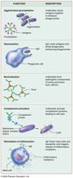
IgG | Main antibody in serum; crosses placenta; opsonization, neutralization, complement activation |
IgA | Secretions (tears, saliva, milk); agglutination, neutralization |
IgM | First antibody secreted; potent agglutination and complement activation |
IgE | Allergic responses; binds mast cells and basophils |
IgD | Antigen receptor on B cells |
Conclusion
The lymphatic and immune systems are essential for fluid balance, fat absorption, and defense against pathogens. Understanding their structure, function, and disorders is critical for diagnosing and treating immune-related diseases.
