 뒤로
뒤로Tissues and the Integumentary System: Structure, Function, and Histology
스터디 가이드 - 스마트 노트
 자료에 맞춘 맞춤형 노트, 핵심 정의, 예시, 맥락을 확장해 제공합니다.
Chap. 6: Tissue Classification
Hierarchy of Organization in the Human Body
The human body is organized into a hierarchy of structural levels, each with distinct roles in maintaining life and function.
Cell: The basic functional unit of living organisms.
Tissue: A group of cells with similar structure and function.
Organ: Composed of multiple tissue types working together for a specific function.
Organ System: A group of related organs performing a common function.
Understanding tissue histology provides insight into the structural basis of organ physiology, as the cellular composition of a tissue or organ often determines its function.
Epithelial Tissue
Characteristics of Epithelial Tissue
Epithelial tissues cover body surfaces, line cavities, and form glands. They are specialized for protection, absorption, filtration, excretion, secretion, and sensory reception.
Cellularity & Specialized Contacts: Composed almost entirely of tightly packed cells with specialized junctions.
Polarity: Exhibits an apical (top) surface and a basal (bottom) surface.
Supported by Connective Tissue: Attached to a basement membrane.
Avascularity: Lacks blood vessels; nutrients diffuse from underlying tissues.
Regeneration: High capacity for renewal due to frequent cell loss from friction.
Classification of Epithelia
Epithelia are classified based on the number of cell layers and the shape of the cells.
Number of Layers: Simple (one layer) or Stratified (multiple layers).
Cell Shape: Squamous (flat), Cuboidal (cube-shaped), Columnar (tall and column-like).
Note: Glands are formed by epithelial cells.
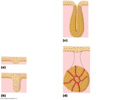
Formation of Endocrine and Exocrine Glands
Glands develop from epithelial sheets and are classified as:
Exocrine Glands: Secrete products into ducts (e.g., sweat, salivary glands).
Endocrine Glands: Release hormones directly into the bloodstream.
Types of Epithelial Tissue
Type | Description | Function | Location |
|---|---|---|---|
Simple Squamous | Single layer of flat cells with disc-shaped nuclei | Diffusion, filtration, secretion | Kidney glomeruli, air sacs of lungs, lining of heart, blood vessels, lymphatic vessels, serosae |
Simple Cuboidal | Single layer of cube-like cells with large, central nuclei | Secretion, absorption | Kidney tubules, ducts and secretory portions of small glands, ovary surface |
Simple Columnar | Single layer of tall cells, some with cilia or goblet cells | Absorption, secretion of mucus/enzymes, ciliary propulsion | Digestive tract, gallbladder, excretory ducts, small bronchi, uterine tubes |
Pseudostratified Columnar | Single layer of cells of varying heights, nuclei at different levels | Secretion (especially mucus), propulsion by cilia | Trachea, upper respiratory tract, male sperm ducts |
Stratified Squamous | Several layers; basal cells cuboidal/columnar, surface cells squamous | Protection from abrasion | Esophagus, mouth, vagina, epidermis of skin |
Stratified Cuboidal | Generally two layers of cube-like cells | Protection | Sweat, mammary, and salivary gland ducts |
Stratified Columnar | Several layers; basal cells cuboidal, superficial cells columnar | Protection, secretion | Male urethra, large ducts of some glands |
Transitional | Resembles both stratified squamous and cuboidal; surface cells dome-shaped | Stretches, permits distension | Ureters, bladder, part of urethra |
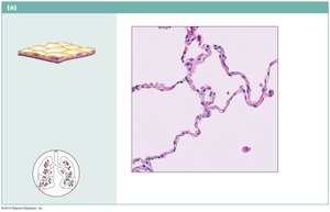
Simple Squamous Epithelium
Description: Single layer of flattened cells with disc-shaped nuclei and sparse cytoplasm.
Function: Allows diffusion and filtration; secretes lubricating substances.
Location: Kidney glomeruli, air sacs of lungs, lining of heart, blood vessels, lymphatic vessels, serosae.
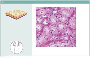
Simple Cuboidal Epithelium
Description: Single layer of cubelike cells with large, spherical central nuclei.
Function: Secretion and absorption.
Location: Kidney tubules, ducts and secretory portions of small glands, ovary surface.
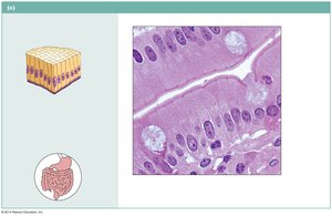
Simple Columnar Epithelium
Description: Single layer of tall cells with round to oval nuclei; may have cilia and goblet cells.
Function: Absorption; secretion of mucus, enzymes; ciliated type propels mucus or reproductive cells.
Location: Digestive tract, gallbladder, excretory ducts, small bronchi, uterine tubes.
Pseudostratified Columnar Epithelium
Description: Single layer of cells of differing heights; nuclei at different levels; may have cilia and goblet cells.
Function: Secretes mucus; propulsion of mucus by ciliary action.
Location: Trachea, upper respiratory tract, male sperm ducts.
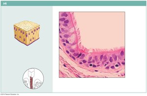
Stratified Squamous Epithelium
Description: Several layers; basal cells cuboidal/columnar, surface cells squamous; keratinized type has dead surface cells full of keratin.
Function: Protects underlying tissues in areas subjected to abrasion.
Location: Esophagus, mouth, vagina (nonkeratinized); epidermis of skin (keratinized).
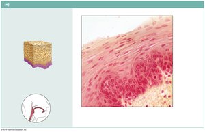
Stratified Cuboidal Epithelium
Description: Generally two layers of cubelike cells.
Function: Protection.
Location: Largest ducts of sweat, mammary, and salivary glands.
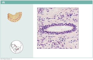
Stratified Columnar Epithelium
Description: Several layers; basal cells usually cuboidal, superficial cells elongated and columnar.
Function: Protection and secretion.
Location: Rare; male urethra, large ducts of some glands.
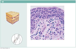
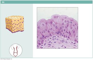
Transitional Epithelium
Description: Resembles both stratified squamous and cuboidal; surface cells dome-shaped or squamouslike depending on stretch.
Function: Stretches readily, permits distension of urinary organs.
Location: Ureters, bladder, part of urethra.
Connective Tissue
Characteristics of Connective Tissue
Connective tissues support, bind, and protect other tissues and organs. They are characterized by:
Rich blood supply (except cartilage, tendons, ligaments).
Many cell types (e.g., fibroblasts, macrophages, adipocytes).
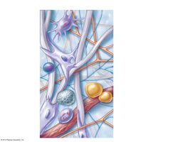
Extracellular matrix (nonliving material between cells):
Ground substance: Interstitial fluid, cell adhesion proteins, proteoglycans.
Fibers: Collagen, elastic, reticular.
Functions of Connective Tissue
Protection of tissues and organs
Support and structural framework
Binding together other tissues
Types of Connective Tissue
Embryonic connective tissue (Mesenchyme): Gives rise to all adult connective tissues.
Connective Tissue Proper:
Loose connective tissue: areolar, adipose, reticular
Dense connective tissue: regular, irregular, elastic
Cartilage: Hyaline, elastic, fibrocartilage
Bone (Osseous tissue)
Blood
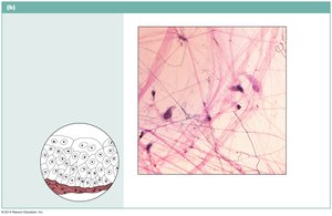
Embryonic Connective Tissue: Mesenchyme
Description: Gel-like ground substance with fibers; star-shaped mesenchymal cells.
Function: Origin of all other connective tissues.
Location: Primarily in embryo.
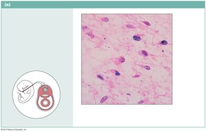
Loose Connective Tissue: Areolar
Description: Gel-like matrix with all three fiber types; cells include fibroblasts, macrophages, mast cells, white blood cells.
Function: Wraps and cushions organs; inflammation; holds tissue fluid.
Location: Under epithelia, packages organs, surrounds capillaries.
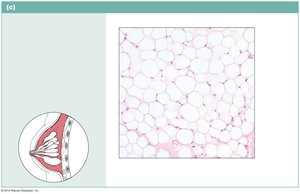
Loose Connective Tissue: Adipose
Description: Sparse matrix; closely packed adipocytes with nucleus pushed to side by fat droplet.
Function: Reserve fuel, insulation, supports/protects organs.
Location: Under skin, around kidneys and eyeballs, abdomen, breasts.
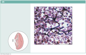
Loose Connective Tissue: Reticular
Description: Network of reticular fibers in loose ground substance; reticular cells on network.
Function: Forms soft internal skeleton (stroma) supporting other cell types.
Location: Lymphoid organs (lymph nodes, bone marrow, spleen).
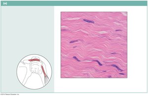
Dense Connective Tissue: Regular
Description: Parallel collagen fibers; few elastic fibers; fibroblast is major cell type.
Function: Attaches muscles to bones or muscles; withstands tensile stress in one direction.
Location: Tendons, ligaments, aponeuroses.
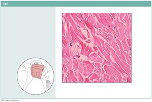
Dense Connective Tissue: Irregular
Description: Irregularly arranged collagen fibers; some elastic fibers; fibroblast is major cell type.
Function: Withstands tension in many directions; structural strength.
Location: Dermis of skin, fibrous capsules of organs and joints, submucosa of digestive tract.
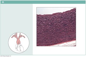
Dense Connective Tissue: Elastic
Description: Dense regular connective tissue with high proportion of elastic fibers.
Function: Allows recoil after stretching; maintains pulsatile flow of blood in arteries.
Location: Walls of large arteries, certain ligaments, bronchial tubes.
Cartilage
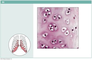
Hyaline Cartilage: Amorphous but firm matrix; supports, reinforces, resists compressive stress. Found in embryonic skeleton, ends of long bones, costal cartilages, nose, trachea, larynx.
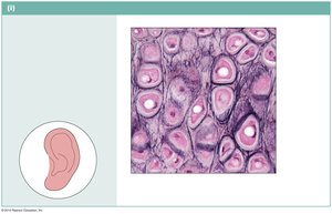
Elastic Cartilage: Similar to hyaline but with more elastic fibers; maintains shape with flexibility. Found in external ear, epiglottis.
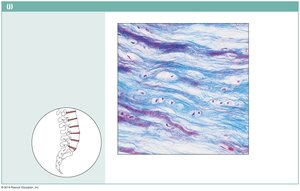
Fibrocartilage: Less firm than hyaline; thick collagen fibers; tensile strength, absorbs shock. Found in intervertebral discs, pubic symphysis, knee joint discs.
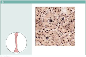
Bone (Osseous Tissue)
Description: Hard, calcified matrix with collagen fibers; osteocytes in lacunae; well vascularized.
Function: Supports, protects, provides levers, stores minerals and fat, site of hematopoiesis.
Location: Bones.
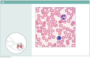
Blood
Description: Red and white blood cells in fluid matrix (plasma).
Function: Transport of gases, nutrients, wastes, and other substances.
Location: Within blood vessels.
Muscle Tissue
Types of Muscle Tissue
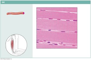
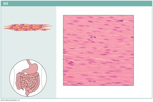
Skeletal Muscle: Voluntary, striated, multinucleate; attached to bones for movement.
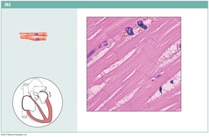
Cardiac Muscle: Involuntary, striated, branching cells with intercalated discs; found in heart walls.
Smooth Muscle: Involuntary, non-striated, spindle-shaped cells; found in walls of hollow organs.
Nervous Tissue
Structure and Function
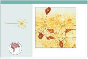
Nervous tissue is specialized for communication via electrical signals. It consists of neurons and supporting cells (neuroglia).
Neurons: Branching cells with long processes (axons, dendrites) for transmitting signals.
Supporting Cells: Protect, support, and insulate neurons.
Location: Brain, spinal cord, nerves.
Chap. 7: The Integumentary System
Functions of the Skin
The skin (cutaneous membrane) is the largest organ of the body and serves multiple vital functions:
Protection against mechanical, chemical, and bacterial damage
Insulation and heat dissipation
Cushioning of body tissues
Prevention of water loss and excretion of wastes
Site for vitamin D synthesis
Skin Structure
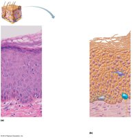
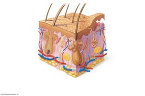
Epidermis: Superficial region with five major layers:
Stratum corneum: Outermost, 20–30 cells thick, bulk of epidermal thickness
Stratum lucidum: Thin, clear layer in thick skin only
Stratum granulosum: Contains granules for waterproofing and support
Stratum spinosum: Several cells thick, capable of division
Stratum basale: Innermost, single cell layer, mitotically active
Dermis: Connective tissue region beneath epidermis; contains two layers:
Papillary layer: Areolar connective tissue, pain and touch receptors
Reticular layer: Dense irregular connective tissue, contains blood vessels, glands, pressure receptors
Hypodermis (subcutaneous tissue): Primarily adipose tissue, not part of skin
Skin Coloration
Skin color is determined by the amount of melanin, carotene, and blood oxygenation. Changes in skin color can indicate various health conditions:
Hypertension: Flushed skin
Anemia: Pale skin
Cyanosis: Bluish skin (asphyxiation/lung disease)
Jaundice: Yellow skin (liver disease)
Addison’s disease: Bronze skin (adrenal cortex hyposecretion)
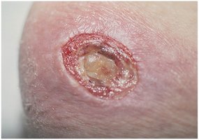
The epidermis lacks blood vessels; nutrients and wastes diffuse from the dermis. Restricted blood supply can cause skin cell death and bedsores (decubitus ulcers), especially in bedridden patients.
Hair and Hair Follicle Structure
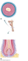
Hair consists of an inner core (medulla), surrounded by a cortex, and covered by a cuticle. The hair root is within the follicle, while the shaft emerges from the scalp. The follicle wall contains several layers, including internal and external epithelial root sheaths and a glassy membrane. Associated structures include sebaceous glands and arrector pili muscles.
Summary Table: Major Tissue Types and Functions
Tissue Type | Main Function | Example Location |
|---|---|---|
Epithelial | Protection, absorption, secretion, filtration | Skin, lining of GI tract, glands |
Connective | Support, binding, protection, transport | Bones, tendons, fat, blood |
Muscle | Movement, posture, heat production | Skeletal muscles, heart, hollow organs |
Nervous | Communication, control, sensation | Brain, spinal cord, nerves |
Additional info: For histology identification, students should be able to recognize tissue types, describe their structure, and relate structure to function and location. Use histology atlases and virtual labs for practice.
