Ch. 1 The Human Body: An Orientation
Marieb - Essentials of Human Anatomy & Physiology 13th Edition
Marieb13th EditionEssentials of Human Anatomy & PhysiologyISBN: 9780135624340당신이 사용하는 게 아니라요?교과서 변경
1장, 문제 17

Mr. Garcia is behaving abnormally, and doctors strongly suspect he has a brain tumor. Which medical imaging device—conventional X-ray, CT, PET, ultrasound, or fMRI—would be best for precisely locating a tumor within the brain? Explain your choice.

검증된 단계별 안내
1
Step 1: Begin by understanding the purpose of the problem, which is to identify the most suitable medical imaging device for precisely locating a brain tumor. Consider the strengths and limitations of each imaging modality mentioned: conventional X-ray, CT, PET, ultrasound, and fMRI.
Step 2: Evaluate conventional X-ray imaging. X-rays are excellent for visualizing dense structures like bones but are not ideal for soft tissue differentiation, such as brain tumors. Therefore, this option is less suitable for the given scenario.
Step 3: Assess CT (Computed Tomography) scans. CT imaging uses X-rays to create detailed cross-sectional images of the body, including soft tissues. It is effective for detecting abnormalities like tumors and provides precise anatomical localization, making it a strong candidate for this case.
Step 4: Consider PET (Positron Emission Tomography) scans. PET imaging is useful for assessing metabolic activity and function, which can help identify active tumor cells. However, it is less precise for anatomical localization compared to CT or fMRI, so it may be used in conjunction with other imaging techniques.
Step 5: Evaluate fMRI (functional Magnetic Resonance Imaging). fMRI is excellent for mapping brain activity and detecting functional changes, but it is not primarily designed for anatomical tumor localization. Ultrasound is generally not used for brain imaging due to the skull's interference with sound waves. Based on this analysis, CT imaging is likely the best choice for precisely locating a brain tumor.

비슷한 문제에 대한 검증된 영상 답변:

이 영상 해법은 위 문제에 도움이 된다고 튜터들이 추천한 것입니다.
영상 길이:
2m

주요 개념

질문에 올바르게 답하기 위해 반드시 이해해야 하는 핵심 개념들은 다음과 같습니다.

Medical Imaging Techniques

Medical imaging techniques are essential tools used to visualize the internal structures of the body. They include various modalities such as X-rays, CT scans, PET scans, ultrasounds, and fMRI. Each technique has its strengths and weaknesses, making them suitable for different diagnostic purposes, particularly in identifying abnormalities like tumors.
추천 영상:
03:24
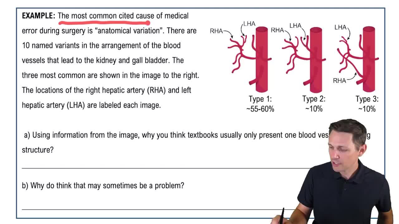
Variation in Anatomy & Physiology Example 1

CT Scan (Computed Tomography)

A CT scan uses X-ray technology to create detailed cross-sectional images of the body, including the brain. It is particularly effective for detecting tumors due to its ability to provide high-resolution images and differentiate between various types of tissues. This makes it a preferred choice for initial assessments of suspected brain tumors.
추천 영상:
가이드 코스
6:42
Map of the Lesson on Innate Immunity

fMRI (Functional Magnetic Resonance Imaging)

fMRI is a specialized type of MRI that measures brain activity by detecting changes in blood flow. While it is excellent for understanding brain function and mapping active areas, it is less effective for locating tumors compared to CT scans. Thus, while fMRI provides valuable information about brain activity, it is not the best choice for tumor localization.
추천 영상:
가이드 코스
06:04
Functional Groups