 뒤로
뒤로A Tour of the Cell: Structure, Function, and Diversity
스터디 가이드 - 스마트 노트
 자료에 맞춘 맞춤형 노트, 핵심 정의, 예시, 맥락을 확장해 제공합니다.
Introduction to Microscopes
Microscopy and the Study of Cells
Microscopes are essential tools in biology, allowing scientists to visualize structures too small for the naked eye. The range of visibility for the human eye, light microscopes, and electron microscopes varies, enabling the study of different biological entities.
Microscope: An optical instrument used to visualize microscopic objects, such as cells.
Light Microscopes: Use visible light to magnify small objects, suitable for most cells and some organelles.
Electron Microscopes: Use electron beams for higher magnification, allowing visualization of subcellular structures.
Example: The size range of atoms, molecules, viruses, bacteria, and cells determines the type of microscope required for observation. 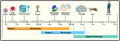
Types of Electron Microscopes
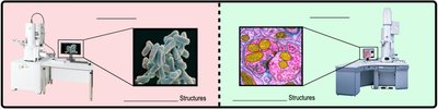
Electron microscopes are divided into two main types, each suited for specific cellular investigations.
Scanning Electron Microscope (SEM): Visualizes external cell surfaces in high detail.
Transmission Electron Microscope (TEM): Visualizes internal cell structures, such as organelles and molecular complexes.
Prokaryotic & Eukaryotic Cells
Domains of Life and Cell Types
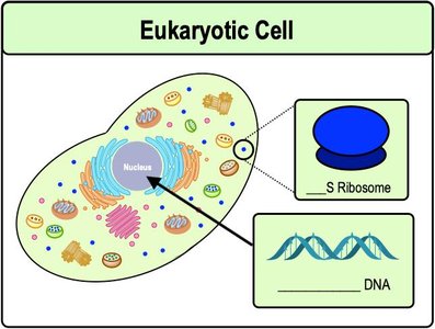
All living organisms are classified into three domains based on cell structure: Bacteria, Archaea, and Eukarya.
Prokaryotic Cells: Lack a nucleus and membrane-bound organelles; include Bacteria and Archaea.
Eukaryotic Cells: Possess a nucleus and membrane-bound organelles; include Eukarya.
Example: Comparison of cell types across domains. 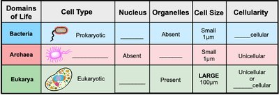
Features of Bacterial Cells
Bacteria are the most abundant and diverse organisms.
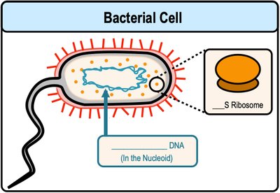
Bacterial DNA: Circular in shape, located in the nucleoid region.
Ribosomes: Small (70S), responsible for protein synthesis.
Cell Division: Binary fission.
Features of Eukaryotic Cells
Eukaryotic cells are characterized by their complexity and presence of organelles.
Nucleus: Contains linear DNA and is surrounded by a nuclear envelope.
Ribosomes: Larger (80S), involved in protein synthesis.
Cell Division: Mitosis and cytokinesis.
Prokaryotic vs. Eukaryotic Cells: Key Differences
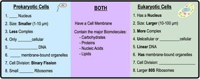
The fundamental differences between prokaryotic and eukaryotic cells are summarized below.
Presence or absence of nucleus and organelles
Cell size and complexity
Type of DNA and ribosomes
Cell division mechanisms
Introduction to Eukaryotic Organelles
Animal vs. Plant Cell Organelles
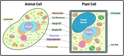
Eukaryotic cells contain several membrane-bound organelles, some of which differ between animal and plant cells.
Animal Cells: Contain lysosomes, mitochondria, and other organelles.
Plant Cells: Contain chloroplasts, central vacuole, and cell wall.
Ribosomes
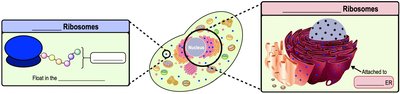
Ribosomes are non-membranous organelles responsible for protein synthesis in all cells.
Translation: The process by which ribosomes build proteins from mRNA.
Free Ribosomes: Float in the cytoplasm.
Bound Ribosomes: Attached to the rough endoplasmic reticulum (ER).
Map of Eukaryotic Organelles
Eukaryotic cell organelles are organized into functional groups, including the endomembrane system, energy organelles, cytoskeleton, and cell junctions. 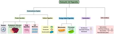
Endomembrane System: Protein Secretion
Components of the Endomembrane System
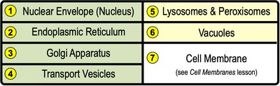
The endomembrane system consists of interconnected membrane-bound organelles that coordinate protein synthesis, modification, and transport.
Nuclear envelope
Endoplasmic reticulum (ER)
Golgi apparatus
Transport vesicles
Lysosomes & peroxisomes
Vacuoles
Cell membrane
Protein Secretion Pathway
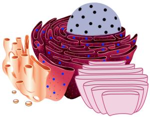
Protein secretion involves several organelles working in sequence.
Nucleus: Stores DNA, which is transcribed into RNA.
Rough ER: Site of protein folding and modification.
Golgi Apparatus: Processes, sorts, and packages proteins for export.
Transport Vesicles: Carry proteins between organelles and to the cell membrane.
Nucleus Structure and Function
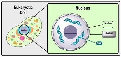
The nucleus is a rounded structure containing most of a cell's genetic material.
Nuclear Envelope: Double membrane surrounding the nucleus.
Nuclear Pores: Allow entry and exit of molecules.
Nucleolus: Site of ribosome assembly.
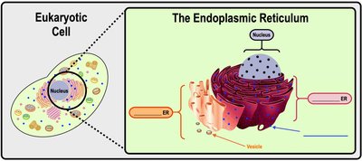
Endoplasmic Reticulum (ER)
The ER is a network of membranous structures continuous with the nuclear envelope.
Rough ER: Ribosome-coated, involved in protein folding and modification.
Smooth ER: Lacks ribosomes, synthesizes lipids, and detoxifies drugs/poisons.
Golgi Apparatus
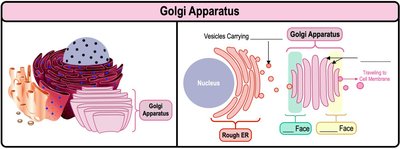
The Golgi apparatus is a stack of flat, membranous sacs functioning as a processing center for proteins and lipids.
Receiving End: "Cis" face.
Shipping End: "Trans" face.
Vesicles from the Golgi can fuse with the cell membrane for secretion.
Endomembrane System: Digestive Organelles
Lysosomes & Peroxisomes
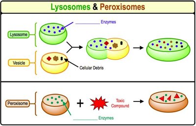
Lysosomes and peroxisomes are specialized vesicles for cellular digestion and detoxification.
Lysosomes: Acidic vesicles containing digestive enzymes, primarily in animal cells.
Peroxisomes: Vesicles containing enzymes that break down toxic compounds and fatty acids.
Central Vacuole
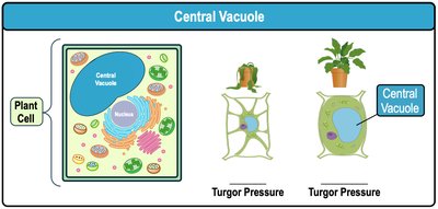
The central vacuole is a large membrane-enclosed vesicle in plant cells, responsible for molecule degradation, recycling, and maintaining turgor pressure.
Occupies most of the cell's volume in plant cells.
Exerts pressure against the cell membrane, supporting plant structure.
Mitochondria & Chloroplasts
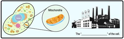
Mitochondria: The Powerhouse of the Cell
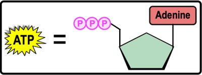
Mitochondria synthesize ATP, the cell's energy currency, through cellular respiration.
ATP (Adenosine Triphosphate): High-energy molecule used to power cellular reactions.
Cellular Respiration: Process of breaking down food sources to produce ATP.
Mitochondria Structure
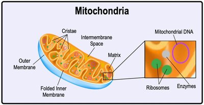
Mitochondria have a unique structure and their own DNA.
Two membranes: outer and folded inner (cristae).
Intermembrane space: region between membranes.
Matrix: contains enzymes, ribosomes, and mitochondrial DNA.
Chloroplasts: Site of Photosynthesis
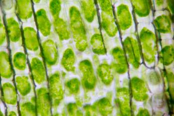
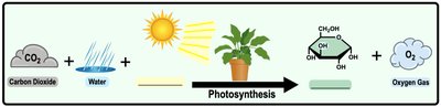
Chloroplasts are green organelles in plant cells where photosynthesis occurs.
Photosynthesis: Uses energy from sunlight to synthesize sugars (glucose).
Products: Oxygen and sugar.
Chloroplast Structure
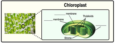
Chloroplasts have two membranes and internal structures for photosynthesis.
Thylakoids: Interconnected disk-shaped sacs.
Grana: Stacks of thylakoids.
Stroma: Innermost region containing enzymes, ribosomes, and chloroplast DNA.
Endosymbiotic Theory
Origin of Mitochondria and Chloroplasts
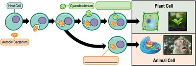
The endosymbiotic theory proposes that mitochondria and chloroplasts originated from independently living prokaryotes engulfed by ancestral eukaryotic cells.
Supporting evidence: Similarities to prokaryotes, such as circular DNA, 70S ribosomes, and binary fission.
Both organelles have double membranes, consistent with engulfment.
Introduction to the Cytoskeleton
Cytoskeleton Components and Functions
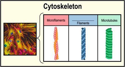
The cytoskeleton is a network of elongated proteins providing cell shape, structure, movement, transport, and biosignaling.
Microfilaments: Small, made of actin proteins.
Intermediate Filaments: Medium-sized, made of variable proteins.
Microtubules: Large, tube-shaped, made of tubulin proteins.
Cilia & Flagella
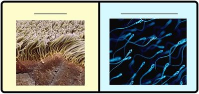
Microtubules form the structural basis of cilia and flagella, which provide cell movement.
Cilia: Multiple hair-like structures moving like oars.
Flagella: Single tail-like structure moving like a whip.
Cell Junctions
Types of Cell Junctions
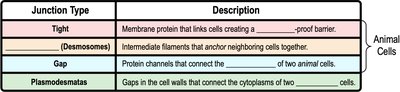
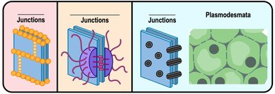
Cell junctions allow neighboring cells to communicate and adhere to one another.
Tight Junctions: Create a waterproof barrier.
Desmosomes: Anchor neighboring cells together.
Gap Junctions: Protein channels connecting animal cells.
Plasmodesmata: Gaps in plant cell walls connecting cytoplasms.
 Additional info: These notes expand on brief points with academic context, definitions, and examples to ensure completeness and clarity for General Biology students.
