뒤로
뒤로Cell Division: Mitosis, Meiosis, and Chromosomal Errors
스터디 가이드 - 스마트 노트
 자료에 맞춘 맞춤형 노트, 핵심 정의, 예시, 맥락을 확장해 제공합니다.
Cell Division
Importance of Cell Division
Cell division is fundamental for growth, repair, and reproduction in multicellular organisms. It allows a single cell to develop into a complex organism and enables the replacement of old or damaged cells. - Key Point 1: Most cells, except neurons and gametes, undergo division throughout life. - Key Point 2: Gametes are produced by meiosis, while all other cells divide by mitosis.
Chromosomes and Chromatin
Chromosomes are highly condensed structures of DNA, which is otherwise found in a relaxed form called chromatin. Before cell division, DNA is replicated so each daughter cell receives a complete set. - Key Point 1: Each chromosome consists of two sister chromatids joined at a centromere. - Key Point 2: Chromatin condenses into visible chromosomes during cell division. 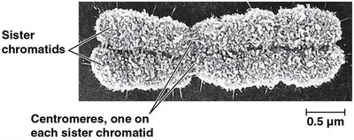
The Cell Cycle
Phases of the Cell Cycle
The cell cycle is divided into several phases that prepare the cell for division. - G1 phase: Cell grows and performs normal functions. - S phase: DNA is replicated. - G2 phase: Cell prepares for division by producing organelles and proteins. - M phase: Mitosis and cytokinesis occur. - Interphase: Includes G1, S, and G2; the cell is not actively dividing. - Checkpoints: Ensure proper progression; failure leads to G0 (cell cycle exit). 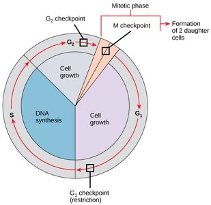
Mitosis
Stages of Mitosis
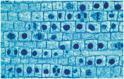
Mitosis is the process by which somatic cells divide for growth, repair, or asexual reproduction. - Prophase: Chromosomes condense, nuclear envelope breaks down, spindle forms. - Metaphase: Chromosomes align at the metaphase plate. - Anaphase: Sister chromatids separate and move to opposite poles. - Telophase: Nuclear envelopes reform, chromosomes decondense. - Cytokinesis: Division of cytoplasm; not a phase of mitosis but follows telophase.
Cytokinesis in Animals vs Plants
Cytokinesis differs between animal and plant cells due to structural differences. - Animal cells: Form a cleavage furrow and contractile ring to pinch in two. - Plant cells: Form a cell plate from Golgi vesicles, which becomes the new cell wall. 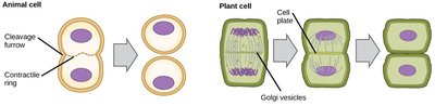
Regulation of Cell Division and Cancer
Control Mechanisms
Cell division is regulated by growth factors and inhibition mechanisms. - Density-dependent inhibition: Cells stop dividing when crowded. - Anchorage-dependent inhibition: Cells require attachment to a substrate to divide.
Cancer and Tumor Formation
Cancer arises when cells bypass normal regulatory mechanisms, leading to uncontrolled growth. - Benign tumors: Remain localized. - Malignant tumors: Invade other tissues and can spread (metastasis). - Metastasis: Cancer cells travel through lymph or blood to establish new tumors. 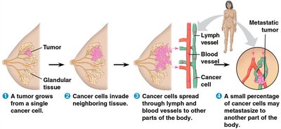
Meiosis
Overview and Significance
Meiosis is the process by which gametes (egg and sperm) are produced, reducing chromosome number by half and enabling genetic diversity. - Diploid (2n): Two sets of chromosomes (somatic cells). - Haploid (n): One set of chromosomes (gametes). - Humans: 22 pairs of autosomes, 1 pair of sex chromosomes.
Phases of Meiosis
Meiosis consists of two sequential divisions: Meiosis I and Meiosis II. - Meiosis I: Homologous chromosomes separate. - Prophase I: Crossing over occurs, exchanging DNA segments for genetic variation. 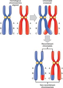 - Metaphase I: Homologous pairs align at the metaphase plate. - Anaphase I: Homologous chromosomes separate. - Telophase I and Cytokinesis: Two haploid cells form. - Meiosis II: Sister chromatids separate, similar to mitosis. - Result: Four non-identical haploid cells.
Comparison: Meiosis vs Mitosis
Meiosis produces four genetically unique haploid cells, while mitosis produces two identical diploid cells. 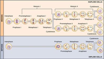
Life Cycle and Genetic Variation
Human Life Cycle
Fertilization combines haploid gametes to form a diploid zygote, which undergoes mitosis and differentiation to develop into an organism. 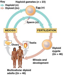
Sources of Genetic Variation
Genetic variation is essential for evolution and adaptation. - Mutations: Original source of variation. - Crossing over: Exchange of DNA during meiosis. - Independent assortment: Random distribution of chromosomes. - Random fertilization: Any sperm can fertilize any egg. - Number of possible gametes: for humans, leading to billions of possible combinations.
Chromosomal Errors
Nondisjunction
Nondisjunction occurs when chromosomes fail to separate properly during meiosis, resulting in gametes with abnormal chromosome numbers. - Consequences: Can lead to miscarriages or genetic disorders. 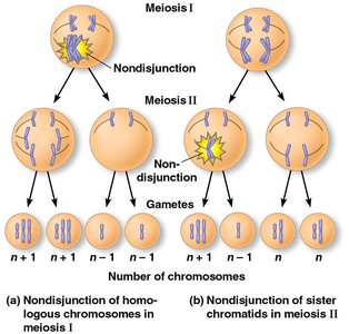
Aneuploidy and Polyploidy
- Aneuploidy: Abnormal number of chromosomes (e.g., monosomy, trisomy). - Polyploidy: Extra sets of chromosomes (common in plants).
Karyotyping
Karyotyping is used to detect chromosomal abnormalities in developing embryos. 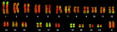
Down Syndrome (Trisomy 21)
Down Syndrome is caused by an extra copy of chromosome 21. - Incidence: 1 in 700 births; risk increases with maternal age. 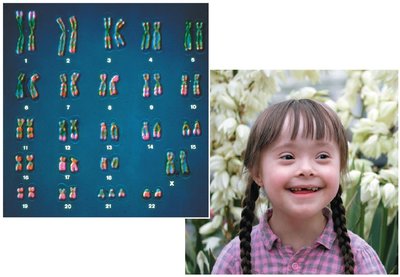
Sex Chromosome Aneuploidy
Sex chromosomes tolerate nondisjunction better than autosomes. - X (Turner syndrome): Only viable monosomy; female is sterile. - XXX: Viable trisomy; possible learning disabilities. - XXY (Klinefelter syndrome): Male with reduced testosterone and sperm. - XYY: Male often taller, possible behavioral issues.
Chromosomal Alterations
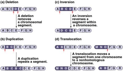
Structural changes in chromosomes can cause genetic disorders.
Type | Description | Example |
|---|---|---|
Deletion | Removes a chromosomal segment | Cri du chat (chromosome 5) |
Duplication | Repeats a segment | Can cause developmental disorders |
Inversion | Reverses a segment within a chromosome | May disrupt gene function |
Translocation | Moves a segment to a nonhomologous chromosome | Chronic Myelogenous Leukemia (chromosomes 9 and 22) |
Summary Table: Mitosis vs Meiosis
Feature | Mitosis | Meiosis |
|---|---|---|
Purpose | Growth, repair, asexual reproduction | Production of gametes |
Number of divisions | 1 | 2 |
Number of daughter cells | 2 | 4 |
Chromosome number | Diploid (2n) | Haploid (n) |
Genetic variation | None (identical cells) | Yes (unique cells) |
Practice: Identifying Cell Division Phases
Cells can be observed under a microscope to identify the various phases of mitosis and meiosis.